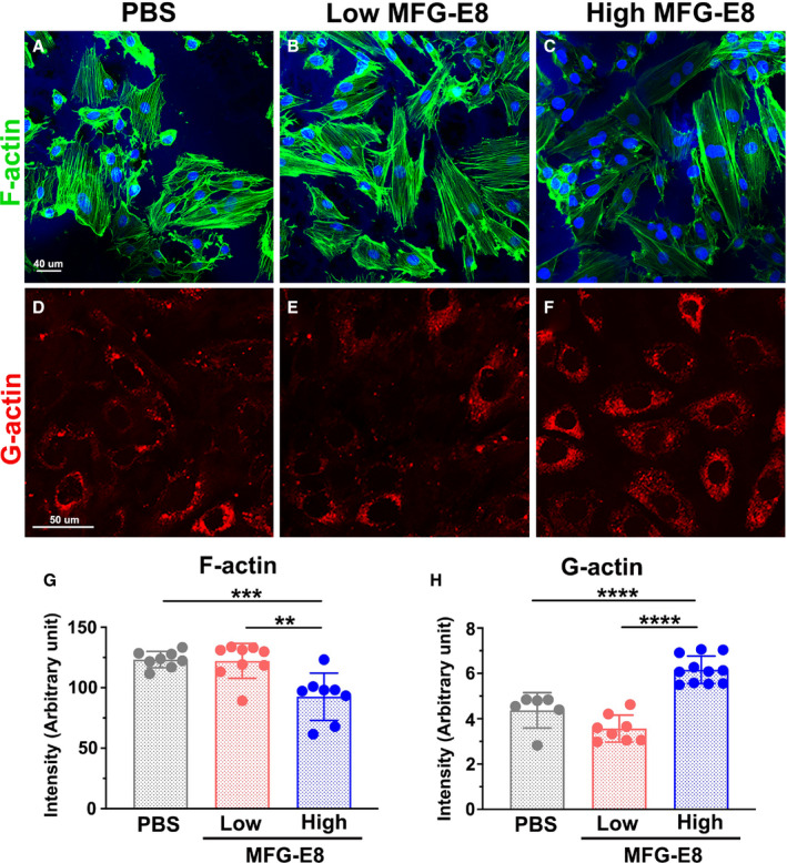
Figure 4. Milk fat globule‐epidermal growth factor (MFG‐E8) modulates actin polymerization in vascular smooth muscle cells (VSMCs) in a dose‐dependent manner.

Representative confocal fluorescent images of phalloidin (filamentous actin [F‐actin], green) A through C, and DNase I (globular actin [G‐actin], red) (D) through (F) staining of A10 VSMCs incubated with low (10 ng/mL) and high (1000 ng/mL) doses of MFG‐E8 for 16 hours. Bar, 50 μm. Three independent experiments were performed, and each experiment was repeated with similar results. In a representative experiment, corresponding quantification of the mean fluorescence intensity of phalloidin for F‐actin (n=8–9) (G) and that of DNase I for G‐actin (n=6–11) H, was performed as described in the Methods section. Data are presented as mean±SD. Each point is derived from an assessment of each picture field. **P<0.01, ***P<0.001, ****P<0.0001, as obtained using 1‐way ANOVA followed by Tukey multiple comparisons test.
