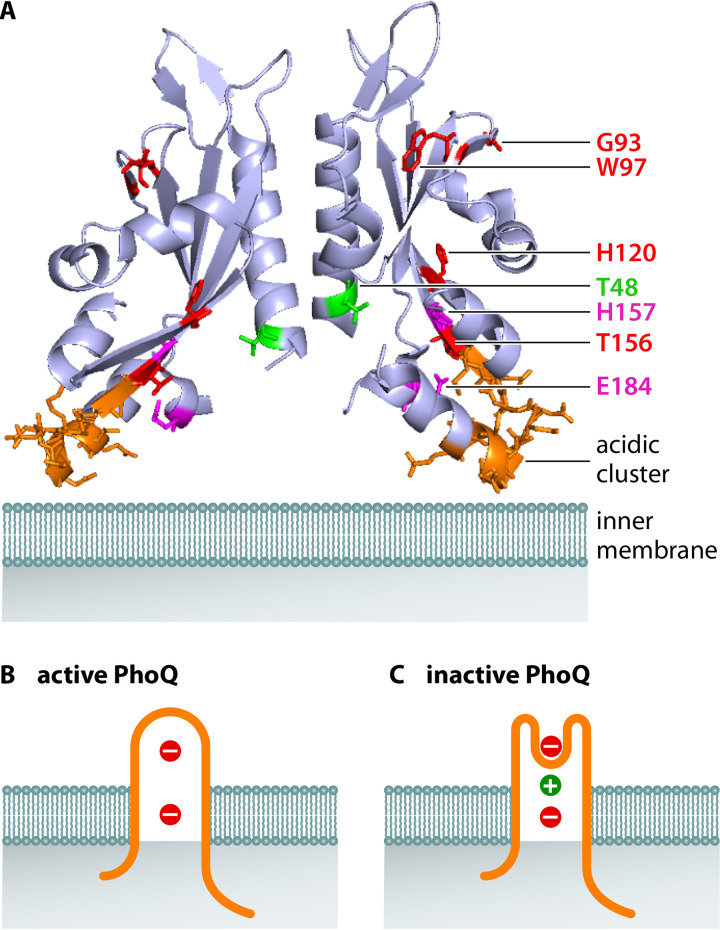
FIG 2.
Structure of PhoQ’s periplasmic domain and model of PhoQ sensing of divalent cations. (A) X-ray structure of the periplasmic domain of the E. coli PhoQ dimer (PDB 3BQ8) (64). The expected position of the inner membrane is in light teal; residues in red correspond to those in which there is direct evidence of a loss of cation binding upon substitution (60); residues in purple correspond to those in which there is indirect evidence in cation sensing through downstream effects on a PhoP-activated gene (62); the residue in green is involved in sensing Ca2+ but not Mg2+ (61); acidic cluster in orange has a minor impact on cation sensing despite being often discussed in the literature (62). (B) In the absence of divalent cations, the membrane negative charges repel the negatively charged periplasmic domain of PhoQ, putting the lever in the “up” position and promoting PhoQ autophosphorylation. PhoQ is in orange, divalent cations in green, and negative charges in the inner membrane in red. (C) When present, the positively charged divalent cations bridge the negative charges in PhoQ and the inner membrane, bringing the lever to the “down” position and hindering PhoQ autophosphorylation.

