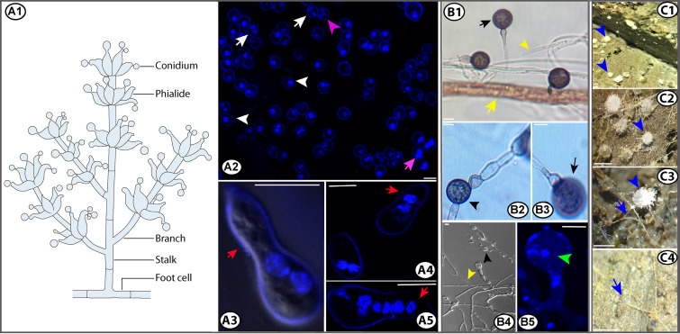
FIG 4.
Schematic diagram of a conidiophore from Morchella, conidial nuclei, and conidial germination (A), chlamydospore formation from conidial hyphae (B), and primordia of morels and probable conidial hyphae at cultivation sites (C). Uninucleate (white arrowheads, A2), binucleate (white arrows, A2), and trinucleate conidia (purple arrowhead, A2). Nucleus mitosis in detached conidia (purple arrow, A2). Conidial germination (red arrows, A3 to A5). Terminal chlamydospores (black arrows, B1 and B3). Intercalary chlamydospores (black arrowheads, B2 and B4). Nuclei of chlamydospores (green arrowhead, B5). Much narrower conidial hyphae (yellow arrowheads, B1 and B4) than other hyphae (yellow arrow, B1). Primordia from morels (blue arrowheads, C1 to C3). Putative conidial hyphae at cultivation sites (blue arrows, C3 to C4). Bars, 5 μm (A and B) or 0.2 mm (C). (Panels A and B are modified from reference 153 with permission from the British Mycological Society.)

