. 2021 Sep 30;10:e71061. doi: 10.7554/eLife.71061

Figure 1. Mutations in MCR reduce aperture number.

(A–E’) Confocal images of auramine O-stained pollen grains from wild-type (Ler) and four mcr EMS mutants. Front (α) and back (α’) show the opposite views of the same pollen grain here and in other figures as indicated. (F, G) 3D reconstructions of tetrad-stage microspores showing lines of INP1-YFP (red arrows) in inp1 and mcr mutants. DP: distal pole. (H, H’) mcr pollen with two apertures. Red boxes mark the regions where apertures are not fused. (I) Percentage of pollen grains with indicated number of apertures in pollen populations from mcr, 1n MiMe, and 1n mcr MiMe plants (n = 75–500). Error bars represent SD, calculated from 4 to 6 independent biological replicates. Apertures are indicated with arrowheads in (A–E’) and (H, H’). Scale bars, 10 μm.

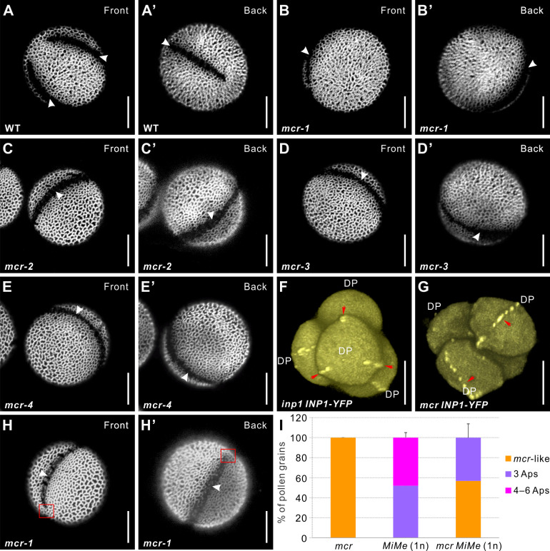
Figure 1.

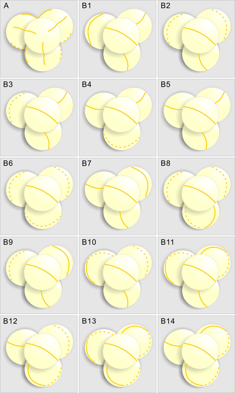
Figure 1—figure supplement 1. Diagrams summarizing the INP1-YFP localization in inp1 and mcr tetrads, based on confocal imaging and 3D reconstruction of DMC1pr:INP1-YFP-expressing tetrads.

Figure 1—figure supplement 1.

(A) Positions of three equidistant lines formed by INP1-YFP in tetrad-stage inp1 microspores always appear coordinated between the sister microspores, with each line in one microspore facing a line in one of its sisters. (B1-B14) Examples of placement of INP1-YFP ring-shaped lines in 14 mcr tetrads, which suggest that the lines in sister microspores are positioned independently. In all tetrads, the INP1-YFP lines in front-facing microspores (with the polar axis perpendicular to the plane of image) were oriented the same way to compare the positioning of the lines in three sister microspores between the tetrads. Solid lines and dotted lines represent the INP1-YFP lines that are, respectively, visible and invisible in that view.

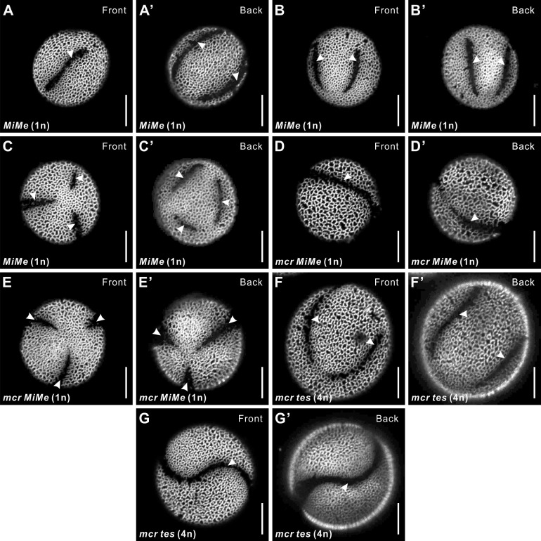
Figure 1—figure supplement 2. The reducing effect of mcr mutations on aperture number is manifested across different ploidy levels and arrangements of microspores.

Figure 1—figure supplement 2.

(A–C’) Representative images of 1n MiMe pollen with three apertures (A, A’), four apertures (B, B’), and six apertures (C, C’). (D–E’) Representative images of 1n mcr MiMe pollen with mcr-like aperture (D, D’) and three apertures (E, E’). (F–G’) Representative images of 4n mcr tes pollen with four apertures (F, F’) and fused apertures (G, G’). Apertures are indicated with arrowheads. Scale bars, 10 μm.