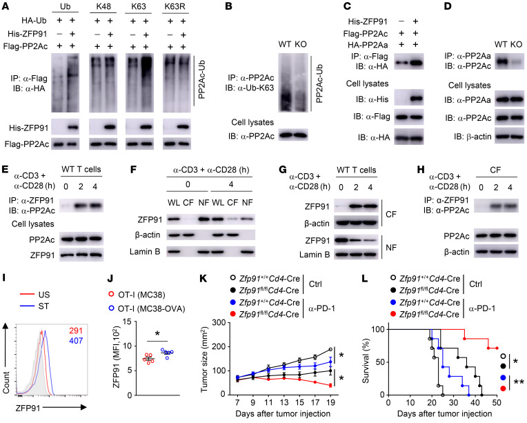
Figure 8. ZFP91 enforces PP2A holoenzyme assembly.
(A) IB analysis of ubiquitinated PP2Ac in HEK293T cells transfected with the indicated vectors. (B) IB analysis of PP2Ac K63-linked ubiquitination in Zfp91+/+ Cd4-Cre (WT) and Zfp91fl/fl Cd4-Cre (KO) CD90.2+ T cells stimulated with anti-CD3 and anti-CD28 antibodies for 4 hours. (C) Lysates from HEK293T cells transfected with the indicated vectors were subjected to IP. (D) Lysates from WT and KO CD90.2+ T cells stimulated with anti-CD3 and anti-CD28 antibodies for 4 hours were subjected to IP. (E) Lysates from WT CD90.2+ T cells stimulated with anti-CD3 and anti-CD28 antibodies were subjected to IP. (F and G) IB analysis of the indicated proteins in whole-cell lysates (WL), cytoplasmic fractions (CF), and nuclear fractions (NF) of WT CD90.2+ T cells stimulated with anti-CD3 and anti-CD28 antibodies. (H) IB and IP assays using the cytoplasmic fractions of WT T cells stimulated with anti-CD3 and anti-CD28 antibodies. (I) Flow cytometric analysis of ZFP91 level in T cells stimulated with anti-CD3 and anti-CD28 antibodies for 24 hours. Isotype control results are shown in gray shading. US, unstimulated; ST, stimulated. (J) Flow cytometric analysis of ZFP91 expression in OT-I cells in tumors of WT mice given an i.v. injection of 2 × 10⁶ OT-I cells on day 7 after s.c. injection of MC38 cancer cells and MC38-OVA cancer cells (n = 5). (K and L) Tumor growth (K) and survival curves (L) for Zfp91+/+ Cd4-Cre and Zfp91fl/fl Cd4-Cre mice injected s.c. with MC38 cancer cells (n = 7), followed by i.p. injection of anti–PD-1 antibody on days 7, 10, and 13. Ctrl, control antibodies. Data are representative of 3 independent experiments and are presented as the mean ± SEM. *P < 0.05 and **P < 0.01, by 2-tailed Student’s t test (J and K) and log-rank (Mantel-Cox) test (L).

