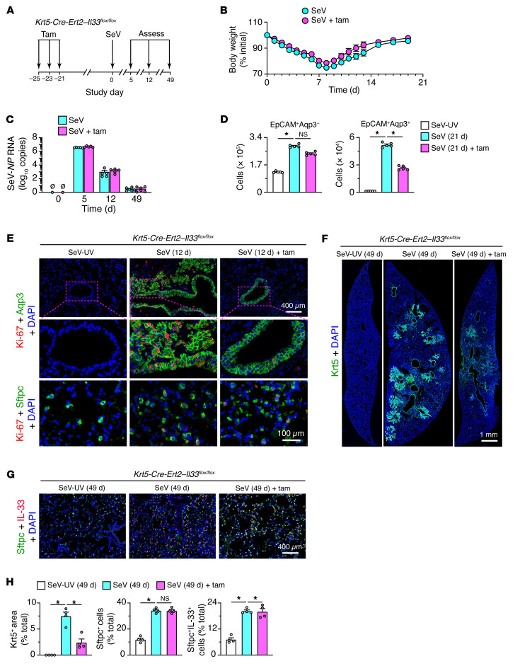
Figure 10. Selective basal ESC depletion in PVLD.
(A) Protocol scheme for analysis of Krt5-Cre-Ert2 Il33fl/fl mice with or without tamoxifen (Tam) treatment followed by SeV infection. (B) Body weights of mice under the conditions in A. (C) Lung levels of SeV-NP RNA in mice under the conditions in A. (D) Cell numbers determined by flow cytometry of lung epithelial cells based on Aqp3 and EpCAM expression after 21 days under the conditions in A. (E) Immunostaining for Ki-67 plus Aqp3 or Sftpc in lung sections from Krt5-Cre-Ert2 Il33fl/fl mice after 12 days under the conditions in A. Scale bars: 400 μm and 100 μm (enlarged insets). (F) Immunostaining for Krt5, 49 days after SeV infection for the conditions in A. Scale bar: 1 mm. (G) Immunostaining for Sftpc and IL-33 under the conditions in F. Scale bar: 400 μm. (H) Quantitation of staining for the conditions in E and F. Data represent results from a single experiment with 4–5 mice per condition, and experiments were replicated twice. *P < 0.05, by ANOVA with Bonferroni correction.

