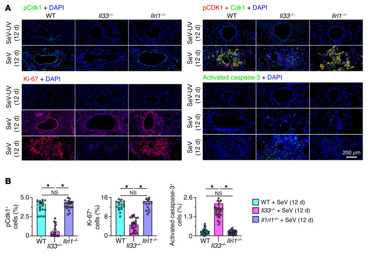
Figure 8. Protein-level validation of IL-33 linkage to cell-cycle activation and progression.
(A) Immunostaining for phosphorylated Cdk1, as well as Cdk1, Ki-67, and activated caspase-3 with DAPI counterstaining of lung sections from mice of the indicated strains 12 days after SeV or SeV-UV infection. Scale bar: 200 μm. (B) Quantitation of the percentage of lung cells that stained positive for the protein markers in A. Data represent results from a single experiment with 5 mice per condition, and experiments were replicated twice. *P < 0.05, by ANOVA with Bonferroni correction.

