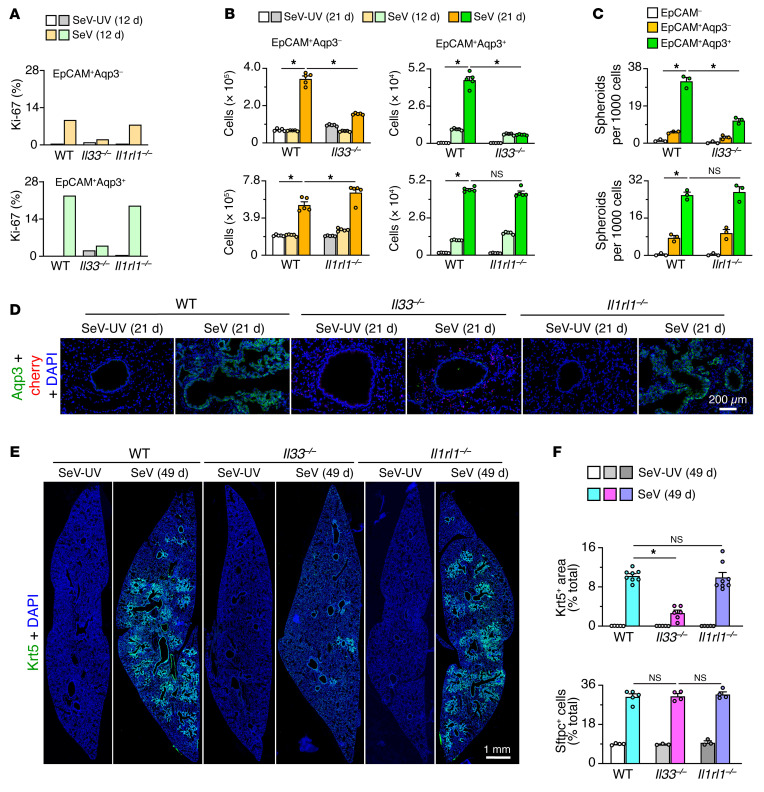
Figure 9. IL-33–dependent basal ESC expansion in PVLD.
(A) Proliferation levels determined by flow cytometry of lung epithelial cells based on Ki-67 expression for WT versus Il33–/– and Il1rl1–/– mice 12 days after SeV versus SeV-UV infection. (B) Corresponding cell numbers from flow cytometry of lung epithelial cells based on Aqp3 and EpCAM expression 12 and 21 days under the conditions in A. (C) Corresponding day-21 lung spheroid formation for FACS-purified cell populations under the conditions in A. (D) Immunostaining for Aqp3 and the IL-33–cherry reporter (cherry) in lung sections under the conditions in A. Scale bar: 200 μm. (E) Immunostaining for Krt5 in lung sections 49 days after SeV or SeV-UV infection under the conditions in A. Scale bar: 1 mm. (F) Quantitation of Krt5 and Sftpc staining under the conditions in E. Data represent results from a single experiment with 3–8 mice per condition, and experiments were replicated twice. *P < 0.05, by ANOVA with Bonferroni correction.

