
Figure 1.
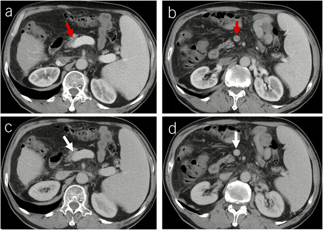
Diagnostic pitfalls of PVT caused by poor-quality CT images. Contrast-enhanced axial CT scans at the portal venous phase showed a mural thrombus occupying the confluence of SMV and splenic vein (red arrow, a) and a complete thrombus occupying the SMV (red arrow, b). Contrast-enhanced axial CT scans at the equilibrium phase showed patent confluence of SMV and splenic vein (white arrow, c) and SMV (white arrow, d). Therefore, mural thrombus shown at portal venous phase may be due to insufficient filling of contrast agent in the confluence of SMV and splenic vein, and complete thrombus may be due to no filling of contrast agent in the SMV. CT, computed tomography; PVT, portal vein thrombosis; SMV, superior mesenteric vein.