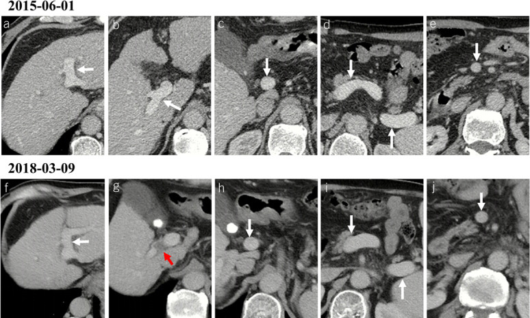
Figure 5.

Development of PVT in a male patient with alcoholic liver cirrhosis. (a–e) Contrast-enhanced axial CT scans on June 1, 2015, showed patent portal venous system vessels, including left portal vein branch (a), right portal vein branch (b), portal vein trunk (c), confluence of SMV and splenic vein (d), splenic vein (d), and SMV (e). The score of each vessel was 0, 0, 0, 0, 0, and 0, respectively. (f–j) Contrast-enhanced axial CT scans on March 9, 2018, showed that de novo thrombus occupied the right portal vein branch (red arrow), while the other portal venous system vessels were still patent. The score of left portal vein branch, right portal vein branch, portal vein trunk, confluence of SMV and splenic vein, splenic vein, and SMV was 0, 2, 0, 0, 0, and 0, respectively. The total PVT score at baseline and during follow-up was 0 and 2, respectively, suggesting the development of PVT. Notes: The red arrows indicate the thrombus, and the white arrows indicate patent vessels. CT, computed tomography; PVT, portal vein thrombosis; SMV, superior mesenteric vein.
