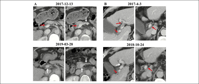
Figure 6.
Recanalization of PVT in liver cirrhosis. (A) Complete recanalization of PVT in a male patient with hepatitis C virus–related cirrhosis. Contrast-enhanced axial CT scans on December 13, 2017, showed a thrombus occupying the portal vein trunk (a) and confluence of SMV and splenic vein (b). Contrast-enhanced axial CT scans on March 28, 2019, showed patent portal venous system vessels (c, d). (B) Partial recanalization of PVT in a male patient with alcoholic liver cirrhosis. Contrast-enhanced axial CT scans on April 3, 2017, showed a thrombus occupying the left portal vein branch (e), right portal vein branch (e), and portal vein trunk (f). Contrast-enhanced axial CT scans on October 24, 2018, showed that the thrombus within the left portal vein branch disappeared, but the previous thrombus remained within the right portal vein branch (g) and portal vein trunk (h). Notes: The red arrows indicate the thrombus, and the white arrows indicate patent vessels. CT, computed tomography; PVT, portal vein thrombosis; SMV, superior mesenteric vein.

