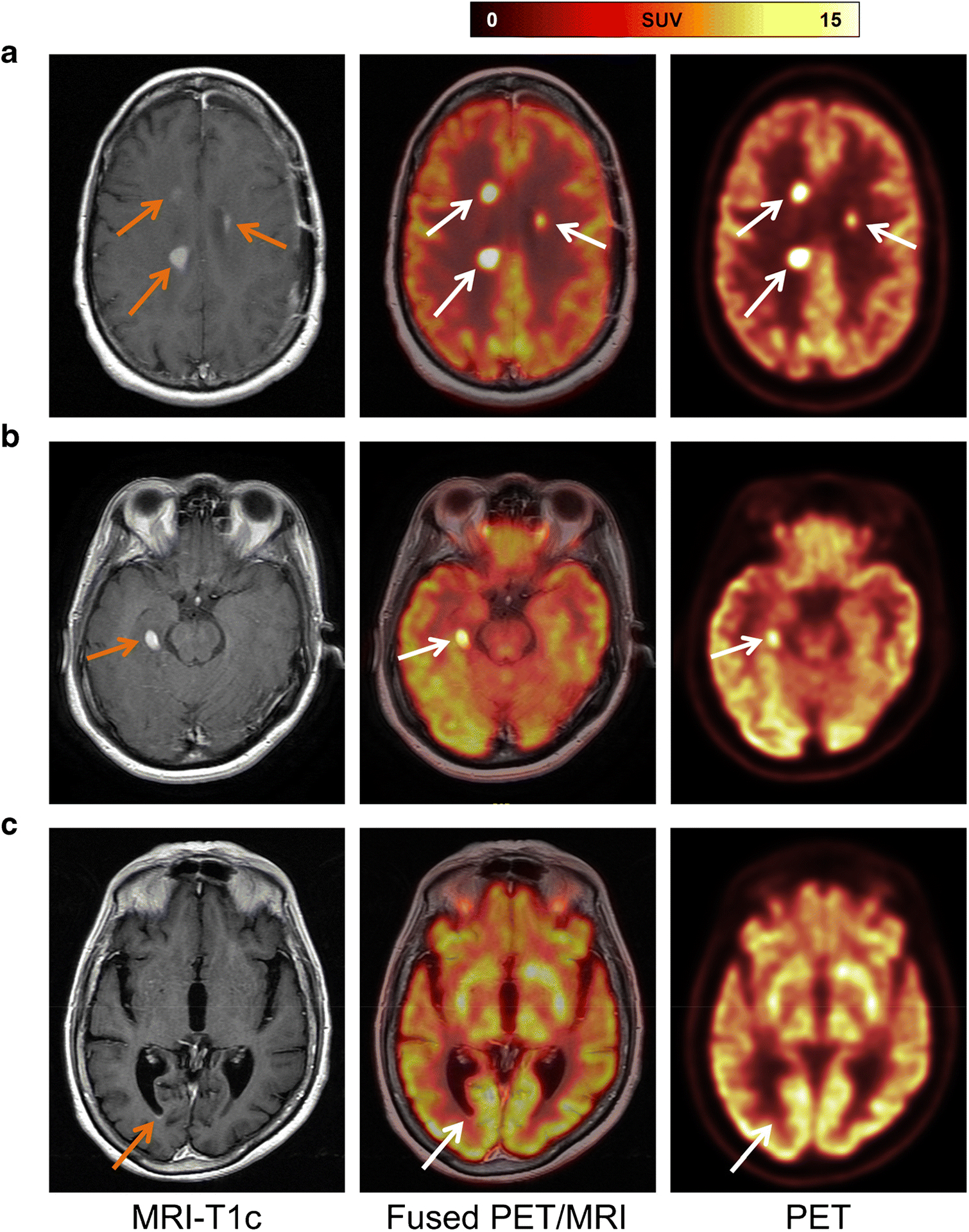
Fig. 2.

Representative cases. (A) Patient diagnosed with secondary CNS lymphoma. Contrast-enhanced T1-weighted MRI shows three enhancing lesions (left, orange arrows), which demonstrate focal [18F]FDG uptake on axial fused PET/MRI (middle, white arrows) and PET images. (B) A representative case of primary CNS lymphoma with a single FDG-avid lesion. Contrast-enhanced T1-weighted MRI depicts an enhancing lesion (left, orange arrow); fused PET/MRI (middle, white arrow) and PET images confirmed focal [18F]FDG uptake. (C) In a patient with primary CNS lymphoma, contrast-enhanced T1-weighted MRI shows a small subependymal enhancement in the right occipital horn (left, orange arrow) that has no correlate on fused PET/MRI (middle, white arrow) or PET images.
