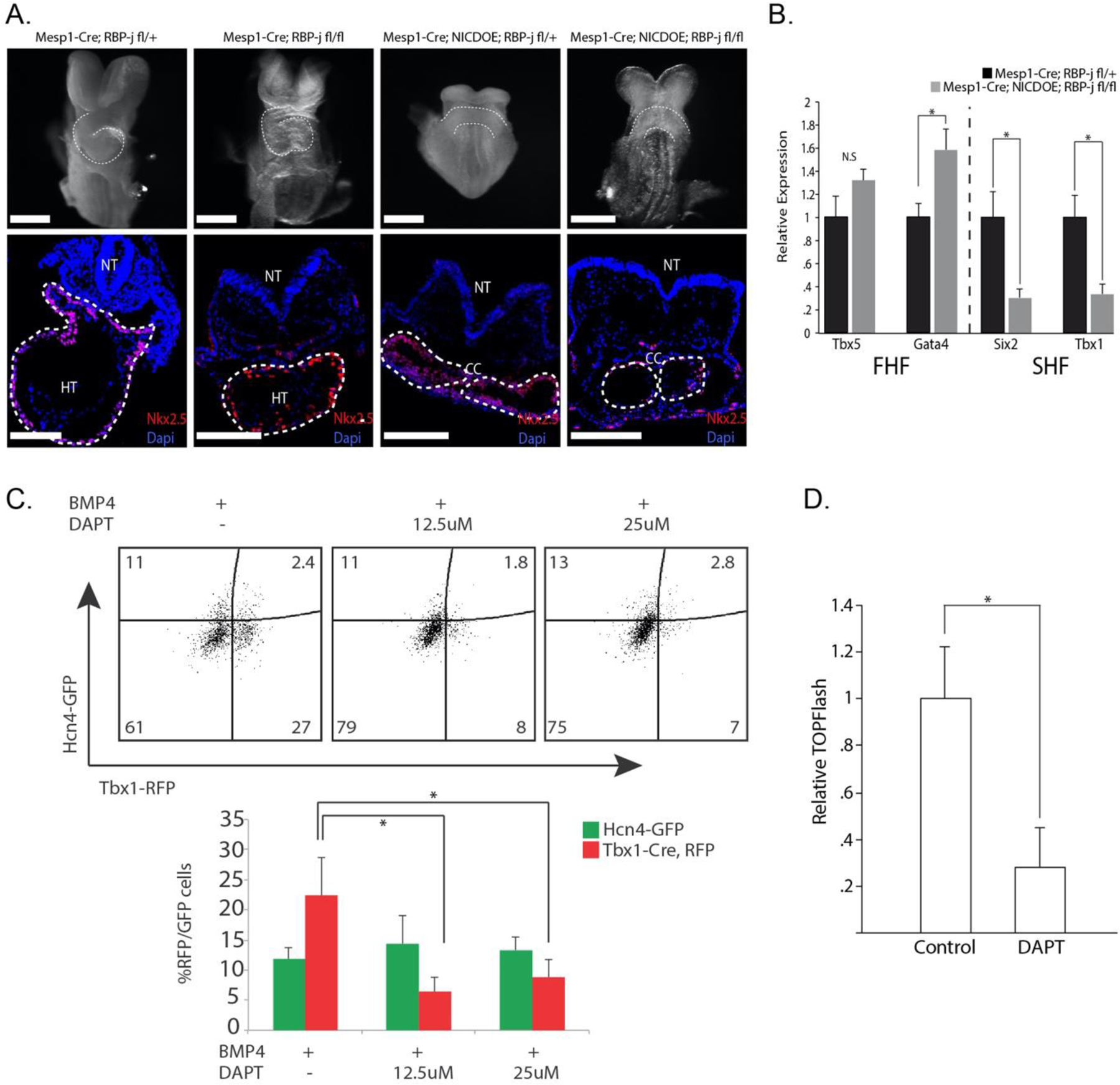
Figure 1. Noncanonical Notch inhibits heart development in Mesp1+ CPCs.

A. E8.5 embryos whole mount and transverse sectioning showing decreased heart size in NICDOE embryos, independent of RBP-j. Cardiac crescent (CC) develops normally, but looping heart (HT) with visible left ventricle (LV) and outflow tract (OT) are not present in mutant embryos (n=10). White scale bars represent 300μm. B. Relative gene expression of FHF/SHF genes from embryos dissected at E8.0. FHF marker Tbx5 shows no significant difference and FHF marker Gata4 shows a slight upregulation in NICDOE embryos. SHF markers Six2 and Tbx1 both show significant downregulation in NICDOE embryos (n=9). C. Representative flow cytometric analysis plots quantifying GFP+/RFP+ cells after DAPT treatment. Percentage of RFP+ (SHF) cells significantly decreases with DAPT treatment. D. DAPT treatment in CPCs during induction leads to decrease in Wnt activity.
