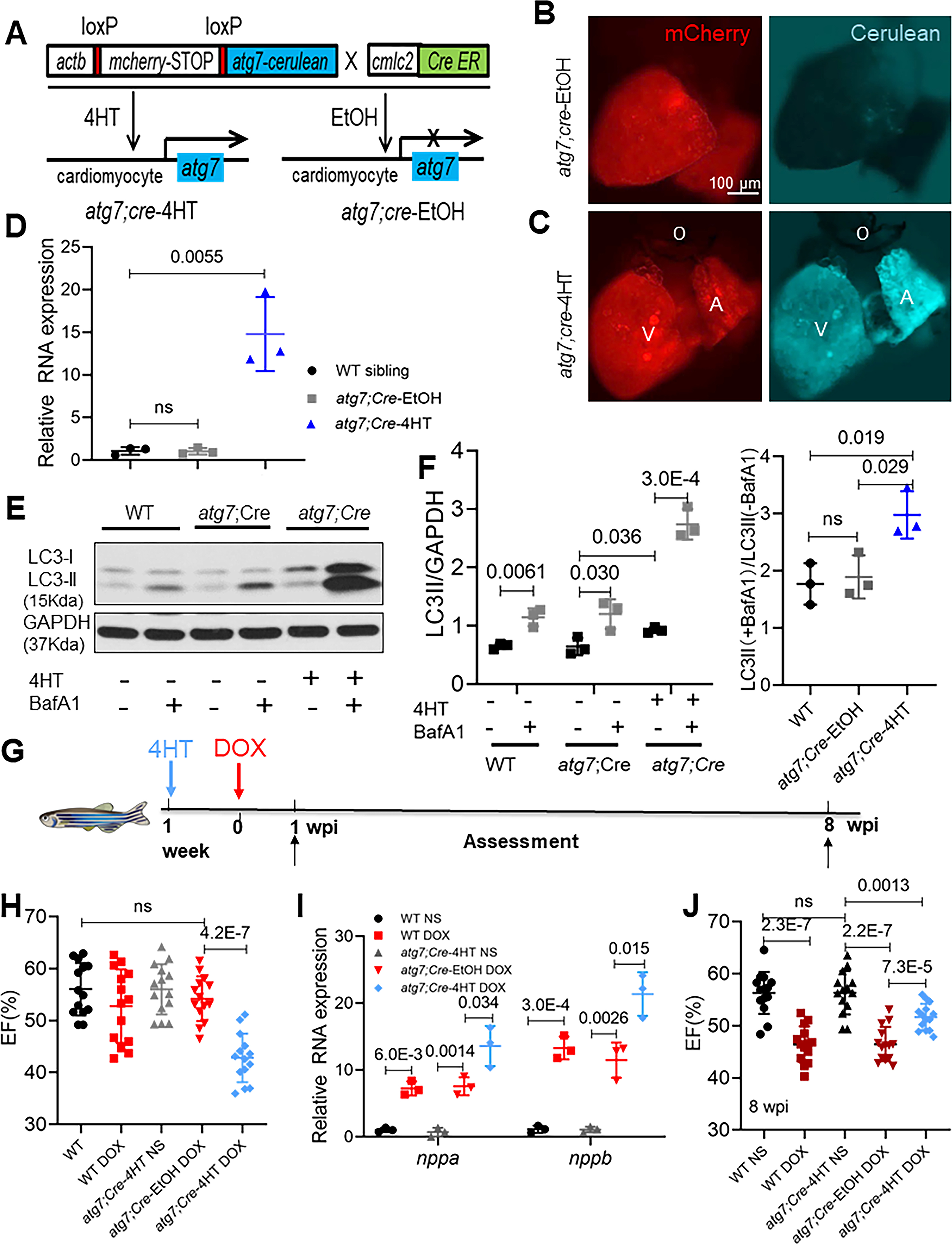
Fig 2. atg7 overexpression (OE) activates autophagy and exerts deleterious effects in the early phase of aAIC.

(A) Schematics of the atg7 conditional transgenic line. (B and C) Fluorescence images of hearts in fish at 1 week after 24-h treatment with 4HT or EtOH. Signals in the cerulean channel represent cardiomyocyte-specific atg7 overexpression after conditional gene activation. (D) Relative transcript level of atg7 RNA in a wild-type sibling and zebrafish with atg7 OE and with and without 4HT treatment. (E) Representative Western blot showing increased LC3-II levels in a zebrafish with atg7 OE and control treated with BafA1 (n=3/treatment). (F) Quantification of LC3-II and the ratio of LC3-II between the hearts treated with and without BafA1 in (E). (G) Schematics of the experimental procedure for activating atg7 in the early phase of aAIC. (H and J) High-frequency echocardiography was performed at the indicated times to quantify cardiac function. One-way ANOVA followed by Tukey’s post hoc test was used. (I) Evaluation of nppa and nppb gene transcript expression by quantitative RT-PCR. (n = 3). Kruskal-Wallis test was used followed by post hoc Tukey’s test in in (D), (F) and (I). WT, wild type; DOX, doxorubicin.
