
Figure 3. Loss of FMRP alters synapse formation and enhances neuronal excitability.
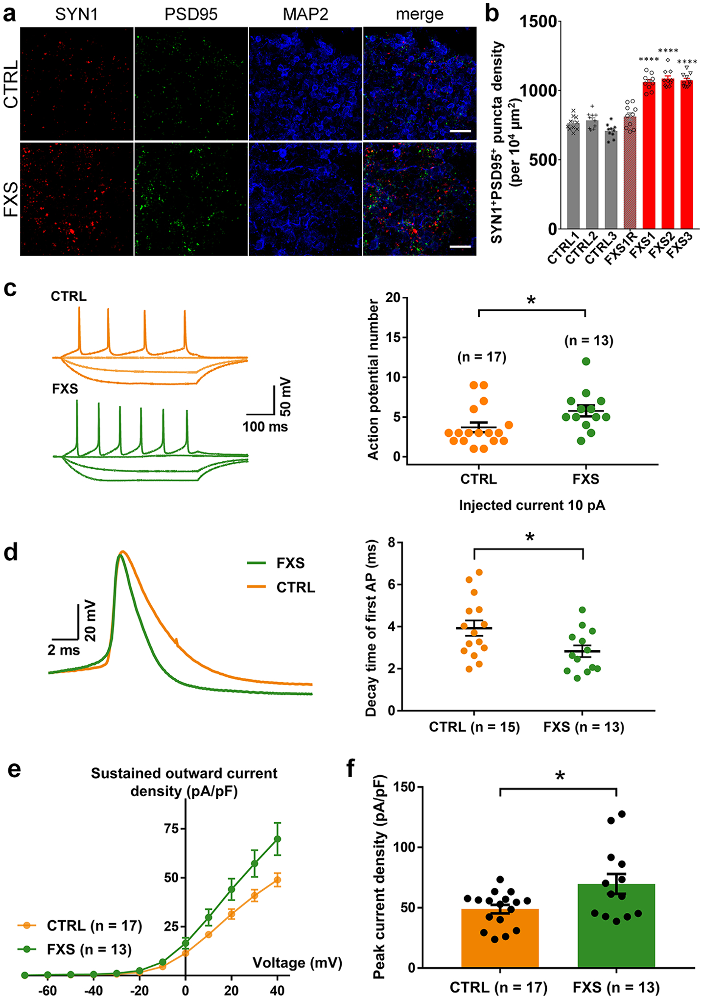
(a-b) Loss of FMRP accelerates synapse formation. Shown are sample images (a) and quantification (b) of SYN1+PSD95+ puncta density in both control and FXS-derived forebrain organoids at day 56. Data are presented as mean ± s.e.m. (n = 10 organoids each line; ****P < 0.0001, one-way ANOVA). Scale bars: 50 μm. (c-f) FXS forebrain organoids exhibit hyperexcitability. Shown in (c) are sample tracings of action potentials (left) and quantification of action potential frequency (right); (d) are sample tracing of the first action potential (left) and quantification of the decay time of first action potential (right); (e,f) are quantification of current-voltage curve (e) and peak current density (f). Cell number (n) recorded and analyzed in each condition is indicated. Data are presented as mean ± s.e.m. (*P = 0.0336 (c), 0.0421 (d), 0.0173 (f), two-tailed unpaired t test or one-way ANOVA).