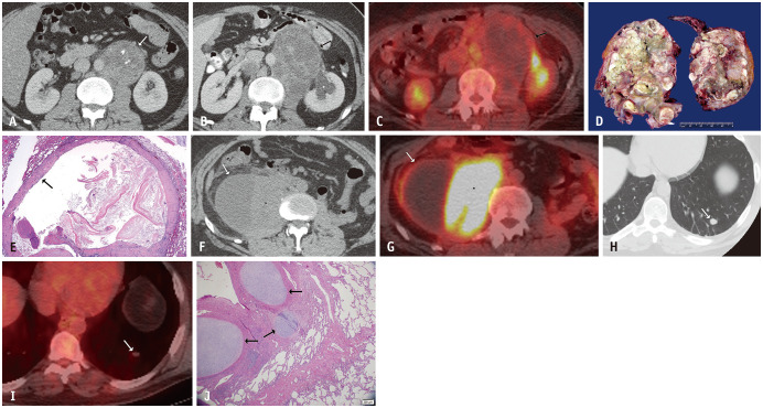
Fig. 3. A 33-year-old male with metastatic testicular non-seminomatous germ cell tumor.
A. Axial contrast-enhanced abdominal CT at the time of presentation showed a solid left retroperitoneal mass with internal calcification (arrow). B. Eight months later, after three cycles of chemotherapy and normalization of tumor markers, an axial contrast-enhanced abdominal CT showed enlargement of the mass with increasing low attenuation predominantly corresponding to necrosis (arrow). The mass also resulted in hydronephrosis of the left kidney (asterisk). C. PET-CT at that time showed no increased FDG-uptake (arrow). D. This mass was resected and gross examination shows near-complete invasion by metastatic mature teratoma with multiple cystic areas filled with yellowish and greenish necrotic material. E. The microscopic appearance of one of the cystic areas (hematoxylin and eosin stain, × 2) shows a squamous epithelial lining (arrow) filled with keratinized squamous epithelial flakes and cellular debris. Findings are consistent with growing teratoma syndrome in the setting of normal tumor markers. F, G. The patient again presented three years later with abdominal pain and normal tumor markers. An axial contrast-enhanced abdominal CT (F) and PET-CT (G) showed a large cystic and solid right retroperitoneal mass (arrows). The solid component demonstrated marked FDG-uptake (asterisks). Pathology was consistent with metastatic teratoma with embryonal carcinoma, which does not produce serum markers. He also had three new lung nodules at that time. H, I. Axial chest CT (H) and PET-CT (I) images showed a left lower lobe nodule with no increased FDG-uptake (arrows). J. The nodules were resected and a histologic image of one of the nodules (hematoxylin and eosin stain, × 40) shows metastatic mature teratoma with cartilaginous differentiation (arrows) within the alveolar parenchyma of the lung (right side of image). FDG = fluorodeoxyglucose

