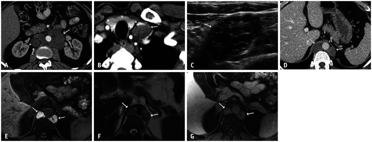
Fig. 4. A 47-year-old male with metastatic testicular non-seminomatous germ cell tumor.
A. After chemotherapy, tumor markers normalized and an axial contrast-enhanced CT of the abdomen showed residual low-attenuation periaortic retroperitoneal lymphadenopathy (arrows). B. An axial contrast-enhanced chest CT image showed a new left supraclavicular lymph node (arrow). C. An ultrasound of the supraclavicular lymph node showed a mixed solid and cystic mass with anechoic spaces (arrow). These masses were resected, and the pathological findings were consistent with metastatic mature teratoma. D-G. Four years later, the patient presented with normal tumor markers and new retrocrural masses (arrows). An axial contrast-enhanced abdominal CT image (D) showed homogeneously low-attenuation masses. A corresponding contrast-enhanced MRI showed marked T1-hyperintensity (E), mild T2-hyperintensity (F), and inherent T1 signal without enhancement on a post-contrast image (G). Note the thin septation within the left retrocrural mass in figure parts (E) and (G) These masses were resected, and the pathological findings demonstrated multicystic masses compatible with metastatic mature teratoma. New left supraclavicular and retrocrural masses with normal tumor markers were consistent with growing teratoma syndrome.

