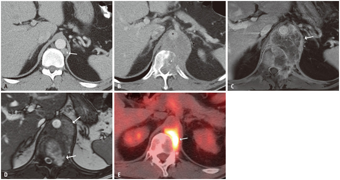
Fig. 5. A 38-year-old male with metastatic testicular non-seminomatous germ cell tumor treated with chemotherapy 6 years earlier.
A. A surveillance axial contrast-enhanced abdominal CT showed a partially calcified left retrocrural lymph node (arrow). No action was taken at that time, and he presented two years later with worsening back pain. B. Axial contrast-enhanced CT showed enlargement of the left retroperitoneal mass (arrow), invading the spine (white asterisk) and encasing and displacing the aorta (black asterisk). C, D. Contrast-enhanced MRI demonstrated peripheral enhancement with enhancing septations (arrow, C) and non-enhancing T2-hyperintensity corresponding to necrosis (arrows, D). E. PET-CT showed fluorodeoxyglucose-uptake along the spine (arrow). The tumor markers were negative, and the pathological findings were consistent with metastatic mature and immature teratomas, which were mostly necrotic with minimal viable malignant tumor.

