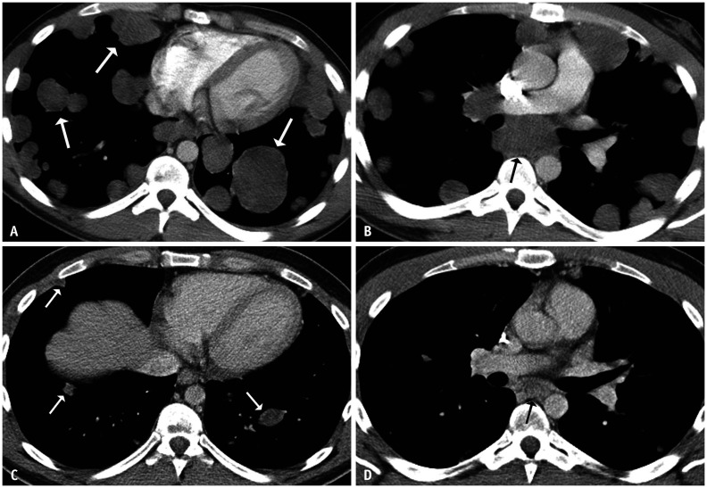
Fig. 6. A 24-year-old male with metastatic testicular non-seminomatous germ cell tumor.
At presentation, there was a large retroperitoneal mass (not shown).
A, B. Axial contrast-enhanced chest CT images showed extensive pulmonary metastatic disease (white arrows) and enlarged mediastinal lymph nodes (black arrow). Following four cycles of chemotherapy, the tumor markers normalized. C, D. A repeat contrast-enhanced CT showed markedly improving pulmonary and mediastinal lesions. The residual lung nodules (white arrows) and a subcarinal lymph node (black arrow) decreased in size and attenuation. Several lung nodules were resected, and the pathological findings were consistent with metastatic mature teratoma with foci of necrosis.

