
FIG. 6.
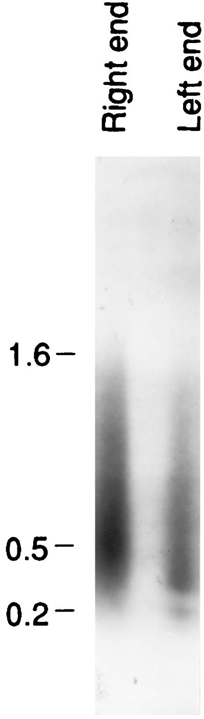
PCRs were carried out with one primer in the telomere for both the right and the left telomere. For the right region a second primer directed to the right, beginning at base 3, was used. For the left region a second primer directed to the left, beginning at base 677, was used. The material was electrophoresed, and the gel was blotted, hybridized with 32P-labeled pXA3, and exposed to film. The result is shown here. Clones were made from this material and sequenced. The sequences indicated that the material consisted of spacer plus telomere sequences as expected. Molecular size markers (in kilobases) are noted at the left.