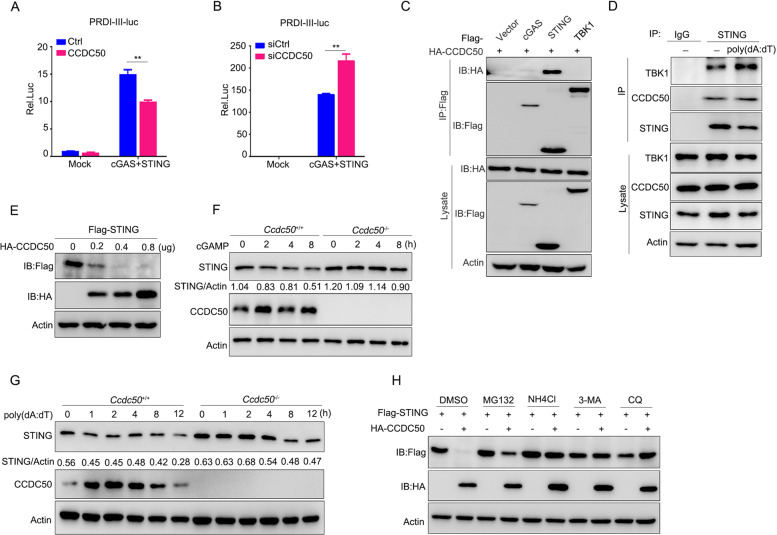
Fig. 4.
CCDC50 associates with STING and targets it for autophagic degradation. A Dual-luciferase activity of PRDI-III-luc in HEK293 cells transfected with plasmids expressing cGAS plus STING along with CCDC50 or empty vector as a control (Ctrl) for 24 h. B Dual-luciferase activity of PRDI-III-luc in HEK293 cells transfected for 24 h with CCDC50 siRNA or nontargeting siRNA (siCtrl) and then transfected with plasmids expressing cGAS plus STING for another 24 h. C Coimmunoprecipitation analysis of the interaction between CCDC50 and cGAS, STING, and TBK1 in cotransfected HEK293 cells. D Immunoprecipitation analysis of the endogenous interaction between CCDC50 and STING in THP-1 cells stimulated with poly(dA:dT) for 4 h. E Immunoblot analysis of lysates from HEK293 cells cotransfected with Flag-STING and an increasing amount of HA-CCDC50. F, G Immunoblot analysis of endogenous STING in Ccdc50+/+ and Ccdc50–/– BMDMs treated with cGAMP (F) and poly(dA:dT) (G) for the indicated time points. H Immunoblot analysis of cell lysates from HEK293 cells cotransfected with Flag-STING and control vector or HA-CCDC50. Fourteen hours after transfection, the cells were treated with DMSO, MG132 (25 μM), NH4Cl (10 mM), 3-MA (5 mM), or CQ (20 μM) for 6 h; actin was used as a loading control. The expression levels of STING and actin were quantitated using ImageJ software. Data are representative of three independent experiments, and error bars are shown as the mean ± SEM (A, B). See also Supplementary Fig. S4. *P < 0.05, **P < 0.01, ***P < 0.001; two-tailed unpaired Student’s t-test

