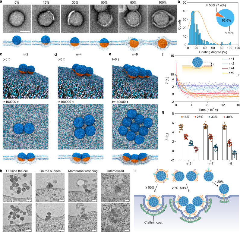
Fig. 4. Endocytic entry mechanism of partially coated NPs.
a Top: TEM images of SiO2 NPs with different cell membrane coating degrees (0%, 15%, 30%, 50%, 80%, and 100%). Scale bar, 50 nm. Bottom: the corresponding final dissipative particle dynamics (DPD) simulation snapshots of the wrapping of the CM-SiO2 NPs by a modeled cell membrane. It shows that the wrapping of a single CM-SiO2 NP with a higher coating degree is easier than that of an NP with a lower coating degree. b Cell membrane coating degree distribution of as-prepared CM-SiO2 NPs, which is calculated from TEM images (n = 325). The inset shows the proportion of SiO2 NPs with a low cell membrane coating degree (<50%). c–e Typical DPD simulation snapshots of multiple CM-SiO2 NPs: aggregation number (n) = 2 (c), 4 (d), and 9 (e). The coating degree of each NP is 33%. The top panel (t = 0 τ) shows the setup of the simulation system and the other panels (t = 160,000 τ) displays the final equilibrated NP-membrane structure at the top view and the profile view. f Time evolution of CM-SiO2 NPs positions along the membrane normal direction. Z represents the distance between the center of the NP and the cellular phospholipid bilayer (inset). g Comparison of positional change of CM-SiO2 NPs with different aggregated numbers (2, 4, and 9) and coating degrees (16%, 25%, 33%, and 40%). Data represents mean ± SD (n = 20). h TEM images showing the different states of CM-SiO2 NPs during receptor-mediated interactions with CT26 cells. Scale bars, 100 nm. i Schematic illustration of a possible endocytic entry mechanism for partially coated NPs.

