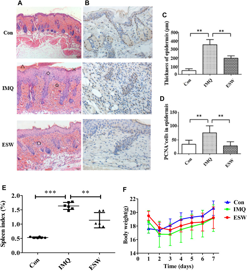
FIGURE 3.
(A) Histological evaluation of skin lesions (H&E staining, magnification ×200); △: microlimb swelling; ×: parakeratosis; ◇: acanthosis; ☆: lymphocyte infiltration; ○: hair follicles and sebaceous glands. (B) Immunohistochemical staining (×200) for proliferating cell nuclear antigen (PCNA) (brown) in mouse back skin. (C,D) Thickness of epidermal and PCNA+ cells of the skin sections of mice from each group on day 7. (E) Histogram of the spleen index of each group. (F) Body weight change in each group from day 1 to day 7. Results were shown as the mean ± SD. **p < 0.01 or ***p < 0.001 vs. IMQ group.

