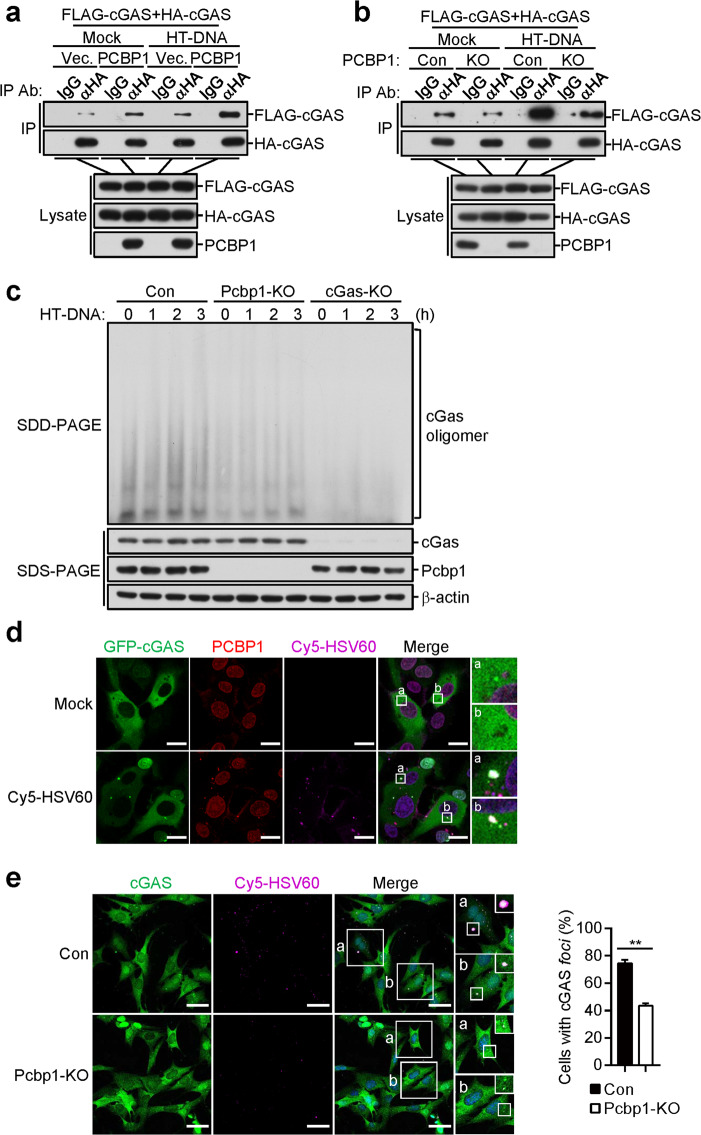
Fig. 6.
PCBP1 is critical for DNA-induced liquid phase separation of cGAS. a PCBP1 promotes the HT-DNA-induced self-association of cGAS in the HEK293 cells. HEK293 cells (1 × 10⁷) were transfected with the indicated plasmids for 20 h. The cells were then mock-transfected or transfected with HT-DNA (10 μg) for 4 h before the coimmunoprecipitation and immunoblot analyses were performed with the indicated antibodies. b Effects of PCBP1 deficiency on the HT-DNA-induced self-association of cGAS in the HEK293 cells. PCBP1-deficient and control HEK293 cells (1 × 10⁷) were transfected with the indicated plasmids for 20 h. The cells were then mock-transfected or transfected with HT-DNA (10 μg) for 4 h before the coimmunoprecipitation and immunoblot analyses were performed with the indicated antibodies. c Effects of PCBP1 deficiency on the HT-DNA-induced oligomerization of cGAS in the MLFs. PCBP1-deficient and control MLFs (1 × 10⁷) were mock-transfected or transfected with HT-DNA (10 μg) for 4 h. Cell lysates were then fractionated by SDD-AGE and SDS-PAGE and analyzed by immunoblotting with the indicated antibodies. d PCBP1 and cGAS form puncta with Cy5-HSV60. HT1080 cells stably expressing GFP-tagged cGAS (2 × 10⁵) were mock-transfected or transfected with Cy5-HSV60 (0.5 μg) for 4 h followed by immunofluorescence staining and analysis. Insets show enlarged images of cGAS-DNA-PCBP1 puncta. Scale bars, 20 μm. e Effects of PCBP1 deficiency on the formation of cGAS-DNA foci in the MLFs. PCBP1-deficient and control MLFs (2 × 10⁵) were mock-transfected or transfected with Cy5-HSV60 (0.5 μg) for 4 h before immunofluorescence staining and analysis. Insets show enlarged images of the foci (left). The percentage of cells with cGAS foci was quantified (right), and at least 100 cells from each group were analyzed. Scale bars, 50 μm. The graph shows the means ± SEM, n = 3. **P < 0.01 (unpaired t-test)

