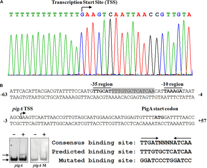
FIGURE 4.
Identification of the Fnr binding site in the pig promoter. (A) Identification of the pig gene transcription start site (TSS) using a 5′-RACE assay (top), and the nucleotide sequence of the pig promoter region (bottom). The PigA start codon, pigA TSS, and the –10 and –35 regions are highlighted in bold. The fnr binding site is shown in shadow. The numbers indicate the distance (shown in nt, – represents upstream, and + represents downstream) to the TSS (+1). (B) Identification of the Fnr binding site. EMSA analysis of the binding between the Fnr and pig promoter probes (pigA) and the mutant pig promoter probe (pigA-M) (Left). Comparison between the conserved Fnr binding motif, predicted Fnr binding sites in the pig promoter, and the mutated nucleotide sequence (Right). Lane –: No protein was added to the reaction mixture. Lane +: 500 nM His6-Fnr was added to the reaction mixture. Each lane contains 0.2 nM-labeled DNA probe. The solid and hollow arrows indicate the free probe and shifted probe, respectively.

