
Figure 5 .
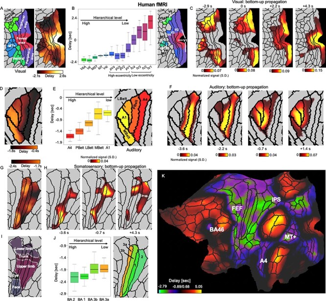
Local embedded propagations within the sensory modalities in human fMRI signals. (A) The local contrast of the PD profile within the 3 visual-related regions defined by a multimodal parcellation atlas (Glasser et al. 2016). (B) The averaged delay values of 13 visual parcels that were arranged according to their hierarchical (Felleman 2009) and retinotopic relationships (Benson et al. 2018). It should be noted that we do not assume any hierarchical relationship between the foveal and peripheral visual parcels. (C) The local trajectory of the bottom-up propagation within the visual system closely follows the PD profile, that is, from the visual association areas to the early peripheral visual areas and then to the early foveal visual regions. (D–F) Results for the auditory system indicated a similar contrast and local propagation between the primary and association auditory areas. (G–I) Results for the somatosensory system show a weaker but still significant relationship between the delay and hierarchical level of 4 somatosensory parcels. The contrast of the PD profile also shows certain correspondence with (J) the somatotopic arrangement (Van Essen and Glasser 2018). (K) The PD profile on a flat brain surface suggests a few other brain regions outside the sensory systems showing large negative delays. Together with the sensory association areas, they compose the task-positive regions that have been shown previously to have strong negative-correlation with the DMN (Fox et al. 2005). Abbreviations: V6A, area V6A; V6, sixth visual area; MT+, MT+ complex; MST, medial superior temporal area; MT/V5, middle temporal area/fifth visual area; V4t, V4 transition zone; V4, fourth visual area (pV4 and fV4 are peripheral and foveal V4, respectively, same thereafter); V3, third visual area; V2, second visual area; V1, primary visual cortex; A4, auditory 4 complex; PBelt, parabelt complex; LBelt, lateral belt complex; MBelt, medial belt complex; BA, Brodmann area; FEF, frontal eye fields; IPS, intraparietal sulcus area.