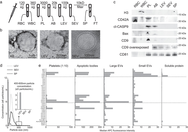
FIGURE 1.

Differential centrifugation of whole blood efficiently separates different cell and vesicle populations. (a) Schematic of the differential centrifugation protocol used to separate red (RBC) and white blood cells (WBC), platelets (PL), apoptotic bodies (AB), large (LEV) and small extracellular vesicles (SEV), as well as soluble protein (SP) and flow through (FT). (b) Representative transmission electron microscopy images of vesicles enriched in AB, LEV and SEV fractions. Corresponding scale bars are inset. (c) Representative Western blots of blood components for histone H3 (H3), CD42A, cleaved Caspase 9 (CASP9), BAX, CD9 and CD81. A CD9 blotted membrane with vesicular and SP components was additionally overexposed. Molecular weight is indicated beside each blot. (d) Nanoparticle tracking analysis data showing the concentration of particles/ml at sizes ranging from 0–1 μm in LEV, SEV and SP fractions. Inset is a quantification of the concentration of particles from 400–600 nm found in each fraction. (d) Median CD9/63/81 fluorescence for 35 capture antibody coated beads from the MACSplex vesicle surface protein flow cytometry assay in the different blood components. The platelet fraction was diluted 1:10 before analysis
