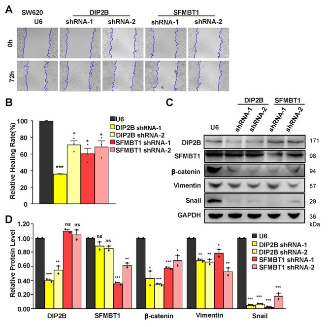
Figure 4 .

Wound healing assays and western blot analysis of the ability of cell migration and invasion in DIP2B and SFMBT1 knockdown cells. (A) Wound healing assays were performed in SW620 cells transfected with U6 and shRNA lentiviruses. Representative images are shown after 0 and 72 h (magnification ×10). The unhealed areas were measured and quantified by ImageJ software. Three independent experiments were performed. (B) Quantification of scratch areas in SW620 cells transfected with U6 and shRNA lentiviruses. Three images were taken for every membrane. (C) Total SW620 cells lysate of transfected cells with U6 (control) and shRNA lentiviruses were analyzed by western blot for the indicated proteins of EMT, DIP2B and SFMBT1. GAPDH was used as a loading control. (D) Quantification of the bands’ intensity in SW620 cells transfected with U6 and shRNA lentiviruses. Two independent experiments were performed. The data are the mean ± SEM; *P < 0.05, **P < 0.01, ***P < 0.001.
