
Figure 5 .
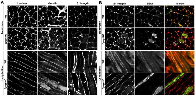
Abnormal localization of focal adhesion complex proteins associated with SPEG deficiency. (A) Transverse (upper panel) and longitudinal (lower panel) WT and Speg-KO TA muscles stained for the laminin, vinculin and β1 integrin. Arrowheads indicate internalized vinculin and arrows indicate internalized β1 integrin, respectively. Scale bar, 20 μm. (B) Transverse (upper panel) and longitudinal (lower panel) TA muscle sections stained for β1 integrin (red) and EEA1 (green). Asterisks indicate the colocalization between internalized β1 integrin and EEA1. Scale bar, 20 μm.