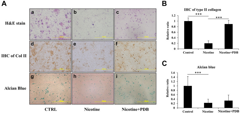
Figure 4.
Chondral regeneration effects of PDB on nicotine-treated 3D neo-cartilage IVD model. We conducted (A) Hematoxylin & eosin (H&E) staining (a-c, upper panel), immunohistochemistry staining of Col II (IHC Col II) (d-f, middle panel) and alcian blue staining (g-i, lower panel) to ascertain the morphologic alterations, collagen and proteoglycan expressions, respectively in control, nicotine-treated and PDB-treated nicotine-IVD group. All the images were captured at 20X magnification (Scale bar: 200 µm). Additionally, these staining signals of (B) Col II and (C) alcian blue were relatively quantified. The results are presented as mean ± S.D. (n = 3; *** P < 0.001).

