
Figure 3.
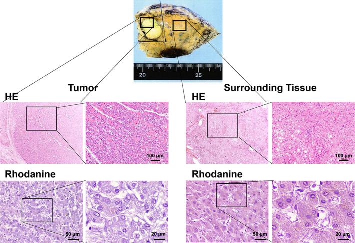
Histological analysis of tumor. A macroscopic of the tumor revealed a 2 cm whitish mass in the liver. The histological analyses revealed a moderately differentiated hepatocellular carcinoma surrounded by cirrhotic tissue with fatty infiltration (Hematoxylin and eosin staining [HE]). Rhodanine staining revealed a slight positively stained area in both tumor and surrounding tissues (black arrows).