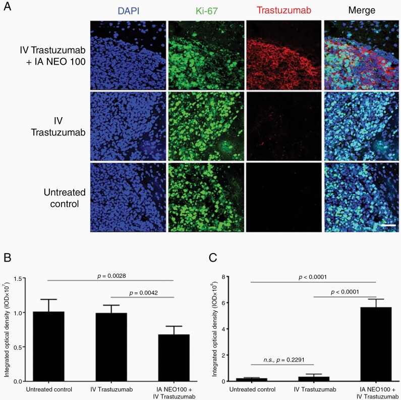
Fig. 2.
Trastuzumab accumulates within tumor tissue, rather than normal brain tissue. Balb/c mice bearing intracranial 4T1-HER2+ cells were treated with intra-arterial (IA) NEO100 (or vehicle), immediately followed by intravenous (IV) trastuzumab, or remained untreated (control). Twenty-four hours later, brains were collected and sections were subjected to immunostaining with fluorescently labeled antibodies recognizing Ki-67 (green) or human IgG (red). DAPI was used as the counterstain (blue). (A) Representative confocal images are shown (scale bar, 50 µm). (B) Quantification of green anti-Ki-67 fluorescence (averages from multiple independent images). (C) Quantification of red anti-human IgG fluorescence (averages from multiple independent images).

