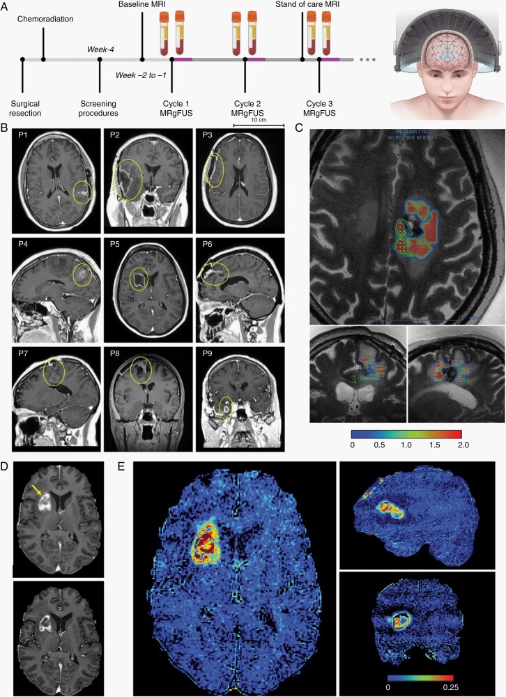
Fig. 1.
Opening of the blood-brain barrier in large brain regions is achieved with transcranial MR-guided focused ultrasound. (A) Schematic diagram of MRgFUS device and study design with BBBO procedures overlapping adjuvant chemotherapy regimen in patients with glioblastoma after surgical resection and concurrent chemoradiation. Blood samples are collected immediately before and after sonications on the same day of the procedure. (B) Representative images of the lesions (yellow circles) on baseline MRI for each patient. (C) Ultrasound can be targeted specifically to an operator-defined contour. The green polygon represents one of many target contours (others not shown). The color map indicates the cavitation dose detected by device upon ultrasound delivery. In (D), the top panel shows contrast extravasation in the areas of increased BBB permeability after MRgFUS for P5 (yellow arrow). The bottom panel shows partial restoration of the BBB integrity approximately 24 hours later, in comparison to the top panel and baseline image of the tumor in (B). (E) Intensity difference maps further show increased BBB permeability in P5 within large regions spatially distributed in the tumor and tumor margins. Heterogeneous changes in contrast extravasation are partially due to the underlying tumor. Abbreviations: BBB, blood-brain barrier; BBBO, BBB opening; MRgFUS, MR-guided focused ultrasound.

