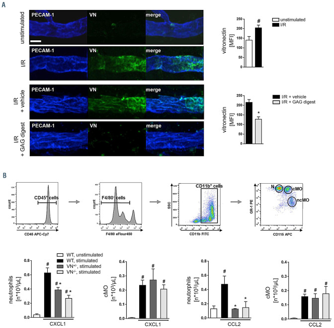
Figure 1.
Role of vitronectin for leukocyte trafficking to inflamed tissue. (A) Using confocal laser scanning microscopy on tissue whole mounts of the cremaster muscle of wil-type (WT) mice, deposition of vitronectin (VN) (green) in the PECAM-1/CD31+ microvasculature (blue) was analyzed, representative images are shown (scale bar: 20 mm). Panels show quantitative results from relative fluorescence intensity measurements for VN in sham-operated WT mice as well as in WT mice undergoing ischemia-reperfusion (I/R) (30/120 min) and intravenous application of glycosaminoglycans digesting enzymes or vehicle (mean±standard error of the mean [SEM] for n=4 animals per group; #P<0.05 vs. sham; *P<0.05 vs. vehicle). (B) Employing multi-channel flow cytometry, the recruitment of neutrophils and classical monocytes to the peritoneal cavity was analyzed 6 hours after intraperitoneal (i.p.) injection of CXCL1 or CCL2, the gating strategy is shown. Panels show results for lipopolysaccharide- treated WT control mice as well as for WT, VN+/-, or VN-/- mice receiving an i.p. injection of CXCL1 or CCL2 (mean±SEM for n=4 animals per group; #P<0.05 vs. control; *P<0.05 vs. WT). cMO: classical monocytes; ncMO: nonclassical monocytes; N: neutrophils; SSC: side scatter.

