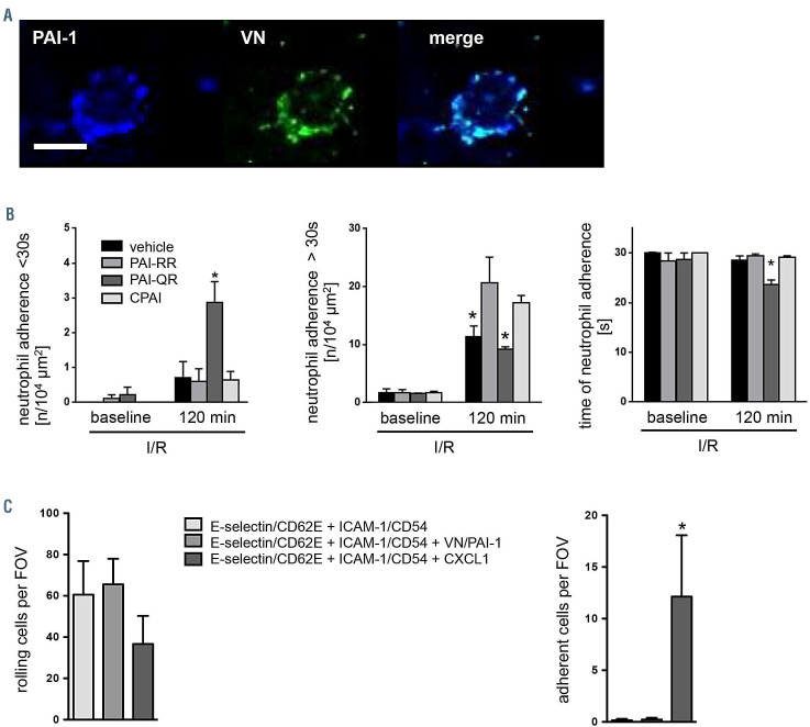
Figure 4.
Effect of vitronectin and plasminogen activator inhibitor-1 heteromers on the stabilization of neutrophil intravascular adhesion. (A) Using confocal laser scanning microscopy on tissue whole mounts of the postischemic cremaster muscle of wild-type (WT) mice, PAI-1 (blue) and vitronectin (VN) (green) deposited on the endothelium of postcapillary venules was detected to co-localize with intravascularly adherent Ly-6G+ neutrophils, representative images are shown (scale bar: 10 mm). (B) Using multi-channel in vivo microscopy on the postischemic mouse cremaster muscle, interactions of Ly-6G+ neutrophils and endothelial cells were analyzed in postcapillary venules. Panels show quantitative results for short adhesion and adhesion time of neutrophils in plasminogen activator inhibitor-1 (PAI-1)-deficient mice receiving vehicle or the PAI-1 mutant proteins PAI-RR (active stable mutant), PAI-QR (unable to bind VN), or CPAI (proteolytically inactive; mean±standard error of the mean [SEM] for n=4 animals per group; *P<0.05 vs. PAI-RR). See also Online Supplementary Table S1. (C) Employing an autoperfused flow chamber assay, intravascular rolling and firm adherence of neutrophils was analyzed in chambers coated with E-selectin/CD62E and ICAM-1/CD54 as well as with or without VN-PAI-1 heteromers or the chemokine CXCL1/KC, panels show quantitative results (mean±SEM for n=7–14 per group; *P<0.05 vs. E-selectin/CD62E + ICAM- 1/CD54 coating). I/R: ischemia-reperfusion; min: muntes, FOV: field of view.

