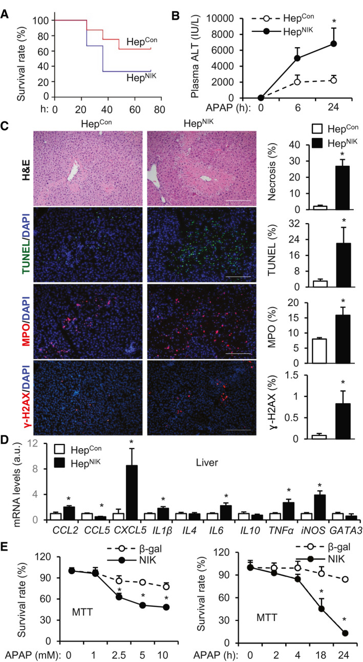
FIG. 2.

HepNIK mice are prone to APAP‐induced acute liver failure. (A) Survival rates following APAP treatment (300 mg/kg body weight). Male HepNIK mice, n = 6; male HepCon mice, n = 8. (B‐D) HepNIK and HepCon male mice were treated with APAP (200 mg/kg). (B) Plasma ALT levels (n = 8 per group). (C) Liver sections were prepared 24 hours after APAP treatment and stained with the indicated agents. Necrotic area was normalized to total area. TUNEL, MPO, or γ‐H2AX cell numbers were normalized to total cell number (n = 3‐4 per group). Scale bar, 200 μm. (D) Liver gene expression 24 hours after APAP treatment (normalized to 36B4 levels, n = 4‐8 per group). (E) Mouse primary hepatocytes were transduced with NIK or β‐gal adenoviral vectors and treated with APAP. Hepatocyte viability was measured by MTT and normalized to baseline levels (n = 6 per group). Data are presented as mean ± SEM. *P < 0.05; (C,D) two‐tailed unpaired Student t test; (B,E) two‐way ANOVA/Bonferroni’s multiple comparisons test.
