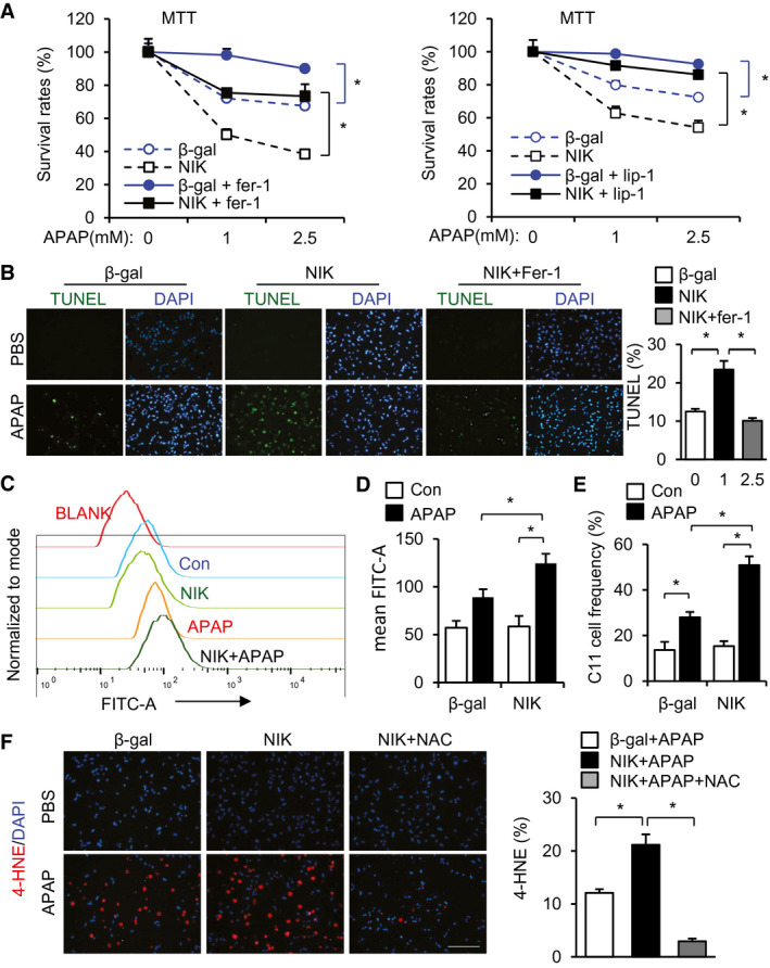
FIG. 7.

NIK enhances ferroptosis of APAP‐treated hepatocytes. (A,B) Mouse primary hepatocytes were transduced with NIK or β‐gal adenoviral vectors, pretreated with 2 μM ferrostatin‐1 or 2 μM liproxstatin‐1, and followed by APAP stimulation for 24 hours. (A) MTT assays (n = 3 per group). (B) TUNEL assays (normalized to total cells, n = 3 per group).( C,D) Mouse primary hepatocytes were transduced with NIK or β‐gal adenoviral vectors, treated with 2.5 mM APAP for 24 hours, stained with BODIPY 581/591 C11 probe, and analyzed by flow cytometry. (C) Representative C11 tracing. (D) Mean C11 density (n = 6 per group). (E) C11high hepatocyte frequency (normalized to total hepatocytes, n = 6 per group). (F) Mouse primary hepatocytes were transduced with NIK or β‐gal adenoviral vectors, treated with APAP for 24 hours in the presence or absence of 500 μM NAC, and stained with anti‐4‐HNE antibody (normalized to total cells, n = 3 per group). Scale bar, 200 μm. Data are presented as mean ± SEM. *P < 0.05; two‐way ANOVA/Bonferroni’s multiple comparisons test. Abbreviations: Fer‐1, ferrostatin‐1; Lip‐1, liproxstatin‐1.
