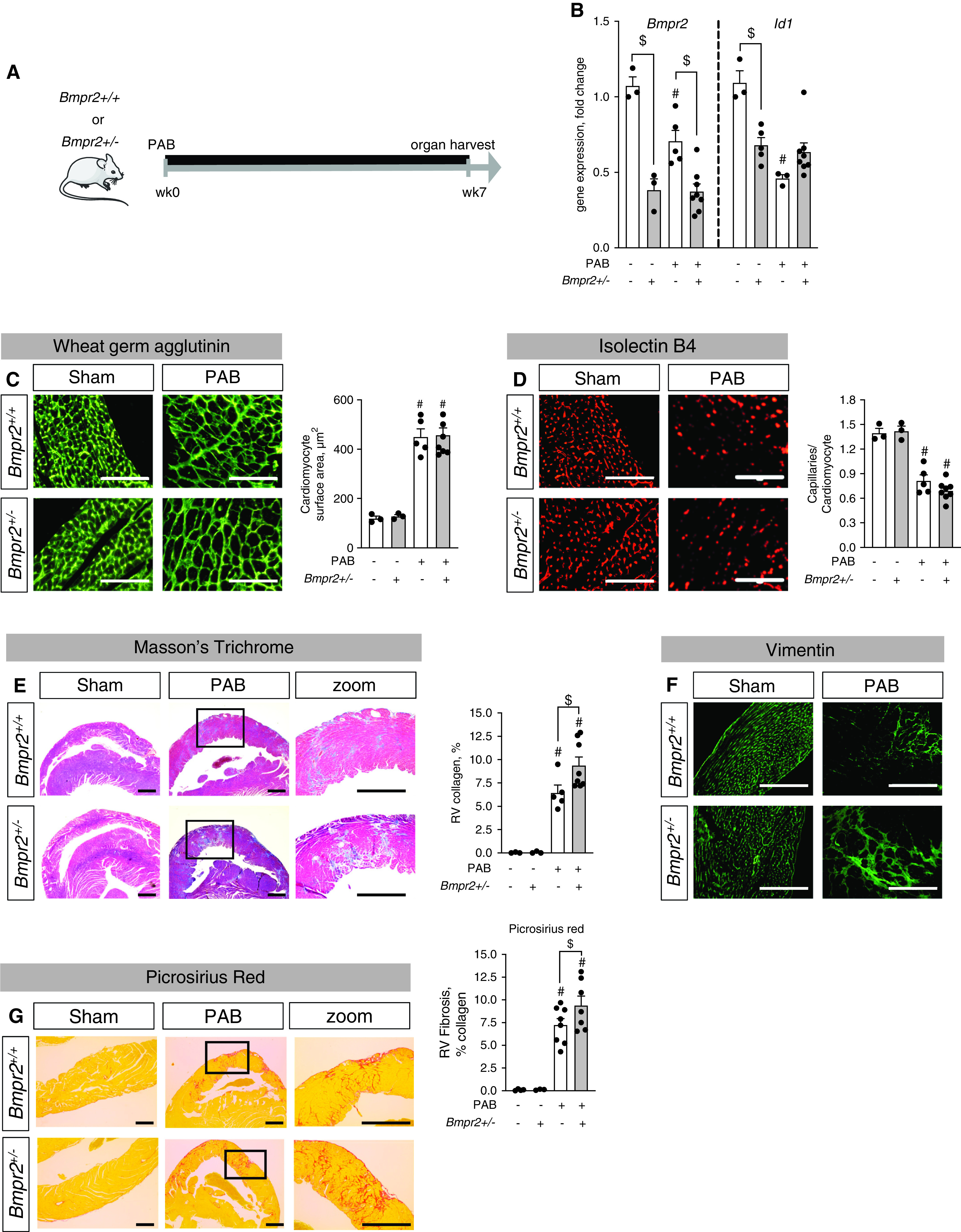
Figure 1.

Low BMP (bone morphogenetic protein) signaling exaggerates right ventricular (RV) fibrosis under pressure overload conditions. (A) Male heterozygous Bmpr2 (BMP receptor type 2) mice (BMPR2+/–) and littermate control mice (BMPR2+/+) underwent sham surgery or moderate pulmonary artery banding (PAB) (around a 24-G needle) for 7 weeks. (B) Gene expression analysis of Bmpr2 and Id1 confirmed reduced BMP signaling in the right ventricle in response to PAB, to concentrations comparable with those of BMPR2+/– mice. (C and D) Histological analyses via staining against wheat germ agglutinin and quantification of cardiomyocyte area demonstrated cellular hypertrophy after PAB independent from BMP signaling (C) together with PAB-induced rarefaction of capillaries as assessed by the ratio of capillaries to cardiomyocytes in isolectin B4 staining (D). (E and F) Masson’s trichrome stain demonstrated collagen accumulation in interstitial and perivascular regions (E) together with increased vimentin+ fibroblasts (F). (G) Picrosirius red stain confirms collagen accumulation in interstitium. n = 3 sham-operated animals; n = 5–8 PAB mice. Two-way ANOVA followed by Tukey’s multiple comparison test. #P < 0.05 versus sham and $P < 0.05 versus BMPR2+/+. Scale bars: C and D, 100 μm; E–G, 200 μm.
