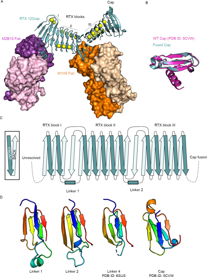
Fig 2. Crystal structure of 123cap bound to the receptor-blocking antibodies M2B10 and M1H5.
(A) Overall structure of 123cap in complex with M2B10 and M1H5 Fabs. Ca2+ ions are shown as yellow spheres. (B) Structural alignment of the capping structure of 123cap with the WT capping structure from the crystal structure of block V (PDB ID: 5CVW), with the WT cap shown in magenta and the fused cap shown in teal. (C) Topology diagram of blocks I–III from 123cap, including the topology of linker 1 and linker 2. (D) Structure of L1, L2, L4 and the region from the capping structure with the same topological motif. Each is colored as an N to C rainbow (blue to red) to show the path of the mainchain.

