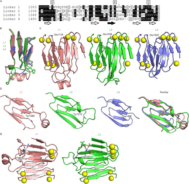
Fig 3. The ACT inter-block linkers are conserved modules.
(A) Sequence alignment of L1, L2, L3, and L4 from B. pertussis ACT. The locations of the core β-sheets are shown below the alignment, with the antiparallel β-strand shown with a black fill. Letters above the alignment denote the subsequent panels that highlight the specified residue. (B) Structural alignment of L1, L2 and L4 (L4 from PDB ID: 6SUS). (C) Conserved Ca2+-binding glutamate at the C-termini of L1, L2, and L4. (D) Conserved core tyrosine/phenylalanine at the N-termini of L1, L2, and L4. (E) Partially conserved buried lysine residue in L1 and L2.

