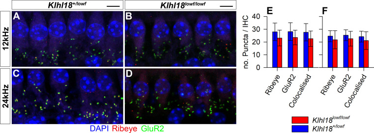
Fig 8. IHC synapses in Klhl18 mutant mice at 6 weeks old.
Representative confocal microscopy images of IHCs at the 12 kHz (A,B) and 24 kHz (C,D) regions of the cochlea from a sample of 7 Klhl18+/lowf mice (A,C), and 7 Klhl18lowf/lowf mice (B,D). A row of IHC nuclei can be seen across the upper portion of panels A-D, stained blue with DAPI. Below this row, clusters of immuno-stained puncta are visible. Green puncta show immuno-positive staining for the post-synaptic receptor subunit GluR2. Red puncta show immuno-positive staining for Ribeye, a protein component of the ribbon synapse apparatus found in inner hair cells. Yellow staining represents overlap between GluR2 and Ribeye (colocalised) staining and is presumed to represent a functional synapse between the inner hair cells and the auditory nerve. Scale bars indicate 5 μm. (E,F) Counts of Ribeye-positive, GluR2-positive and colocalised Ribeye and GluR2-positive puncta are plotted for the 12 kHz region (E) and the 24 kHz region (F). Data are plotted as bars representing the mean count per IHC (error bars indicate the SD) for control (Klhl18+/lowf, blue bars) mice and homozygous mutant mice (Klhl18lowf/lowf, red bars).

