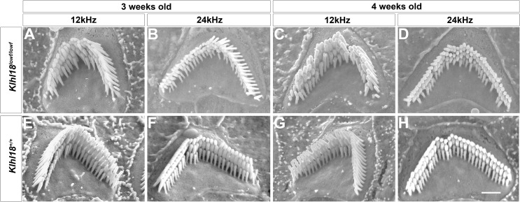
Fig 10. Scanning electron microscopy of typical stereocilia bundles of outer hair cells in Klhl18 mutant mice in mid- and apical turns.
Representative images are shown from (A-D) Klhl18lowf/lowf homozygotes and (E-H) littermate wildtype mice. (A, E) 3 weeks old mice at the 12 kHz best-frequency region; (B, F) 3 weeks old mice at the 24 kHz best-frequency region; (C, G) 4 weeks old mice at the 12 kHz best-frequency region; (D, H) 4 weeks old mice at the 24 kHz best-frequency region. Scale bar (on panel H) represents 1 μm.

