
FIGURE 1.
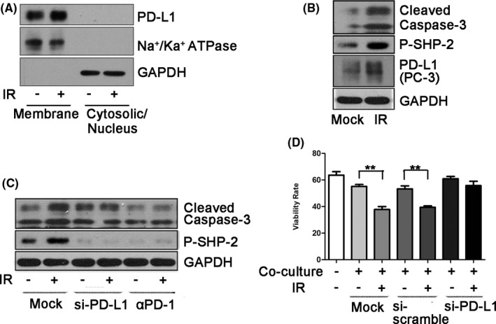
Ionizing radiation induces cancer immunosuppression through upregulation of PD‐L1. A, Cell‐surface expression of PD‐L1 was examined using cell fractionation in MDA‐MB‐231 cells at 2 h after exposure to 5 Gy radiation. Na+/Ka+ ATPase was used as a plasma membrane marker. B, After exposure to 0.1 Gy, PC‐3 cells were co‐cultured with Jurkat cells for 24 h. Cleaved caspase‐3 and pSHP‐2 levels were examined by western blotting (WB) after harvesting Jurkat cells. PD‐L1 was examined after harvesting PC‐3 cells. C, Jurkat cells were co‐cultured with PC‐3 cells treated with 0.1 Gy for 24 h. Cleaved caspase‐3 and pSHP‐2 levels in Jurkat cells were examined by WB after knocking down PD‐L1 expression of PC‐3 cells using siRNA‐PD‐L1 or using an anti‐PD‐1 blocking antibody. The siRNA‐PD‐L1 was transfected into PC‐3 cells for 48 h before co‐culture. D, Flow cytometry was performed to detect annexin V‐FITC/7‐AAD staining to determine the viability of Jurkat cells after co‐culture with PC‐3 cells treated with 0.1 Gy. siRNA‐PD‐L1 or anti‐PD‐1 blocking antibodies were also used in this co‐culture system. Data are presented as the mean ± standard deviation (SD) of triplicate independent experiments. Statistical significance was determined using Student two‐tailed t test. *P < .05, **P < .01