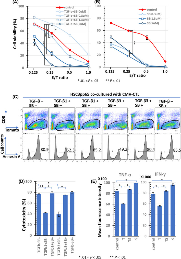
FIGURE 4.

Effect of TGF‐β on cytotoxicity of antigen‐specific CTLs. CMV‐CTLs induced from PBMCs of donor SS were co‐cultured with HSC‐3pp65 in serial E/T ratio for 5 d. HSC‐3pp65 viability was measured using the WST‐1 assay. Cells were treated with SB525234 at the indicated doses in the presence of 10 ng/mL TGF‐β1 (A) or in the absence of TGF‐β1 (B). The mean fluorescence intensity (MFI) of IFN‐γ and TNF‐α concentrations in the supernatant of the CMV‐CTLs co‐cultured with HSC‐3pp65 at an E/T ratio of 1 for 24 h in the presence of SB525334 (1 μmol/L) and/or TGF‐β1 (10 ng/mL) are shown in (C). At 2 d after co‐culture of the CMV‐CTLs and HSC‐3pp65 with TGF‐β1 (10 ng/mL) or TGF‐β3 (10 ng/mL) in the presence or absence of SB525334(1 μmol/L), an annexin V assay was performed. Cells were separated by CD8 and Tomato, the percentage of annexin V+ cells in the Tomato+ gated cells was measured (D), and the cytotoxicity was calculated for each condition (E). The numbers of histograms in panel (D) indicate the percentages of annexin+ cells. SB, SB525334
