
FIGURE 5.
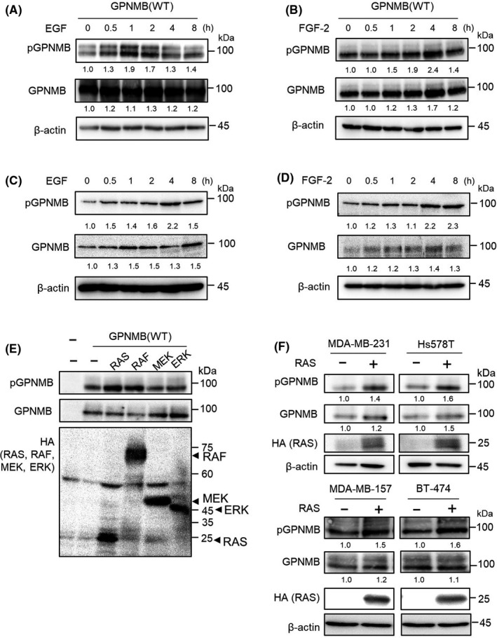
Effect of tyrosine kinase receptor signaling on the serine phosphorylation of glycoprotein non–metastatic melanoma protein B (GPNMB) protein. A,B, 293T cells were transfected with human GPNMB(WT) and treated with 10 ng/mL of EGF (A) or 10 ng/mL of FGF‐2 (B), for the indicated time periods. Immunoblotting for pGPNMB(S530) and total GPNMB was performed. β‐actin was used as a loading control. Intensities of the bands are quantified by Image J and normalized to those intensities of β‐actin. C,D, MDA‐MB‐231 cells were treated with 10 ng/mL of EGF (C) or 10 ng/mL of FGF‐2 (D) for the indicated time periods. Immunoblotting for pGPNMB(S530) and total GPNMB was performed. β‐actin was used as a loading control. Intensities of the bands were quantified by Image J and normalized to those intensities of β‐actin. E, 293T cells were transfected with HA‐tagged RAS (G12V), BRAF (V600E), MEK1 (S218D/S222D), and ERK2 (L73P/S151D). Immunoblotting for pGPNMB (S530), total GPNMB, and HA was performed. F, MDA‐MB‐231, Hs578T, MDA‐MB‐157, and BT‐474 cells were transfected with HA‐RAS (G12V). Immunoblotting for pGPNMB(S530), total GPNMB, and HA was performed. β‐actin was used as a loading control. Intensities of the bands are quantified by Image J and normalized to those intensities of β‐actin