
Figure 5. Serum- and PEMA enveloping-dependent transfection and toxicity of IPs.
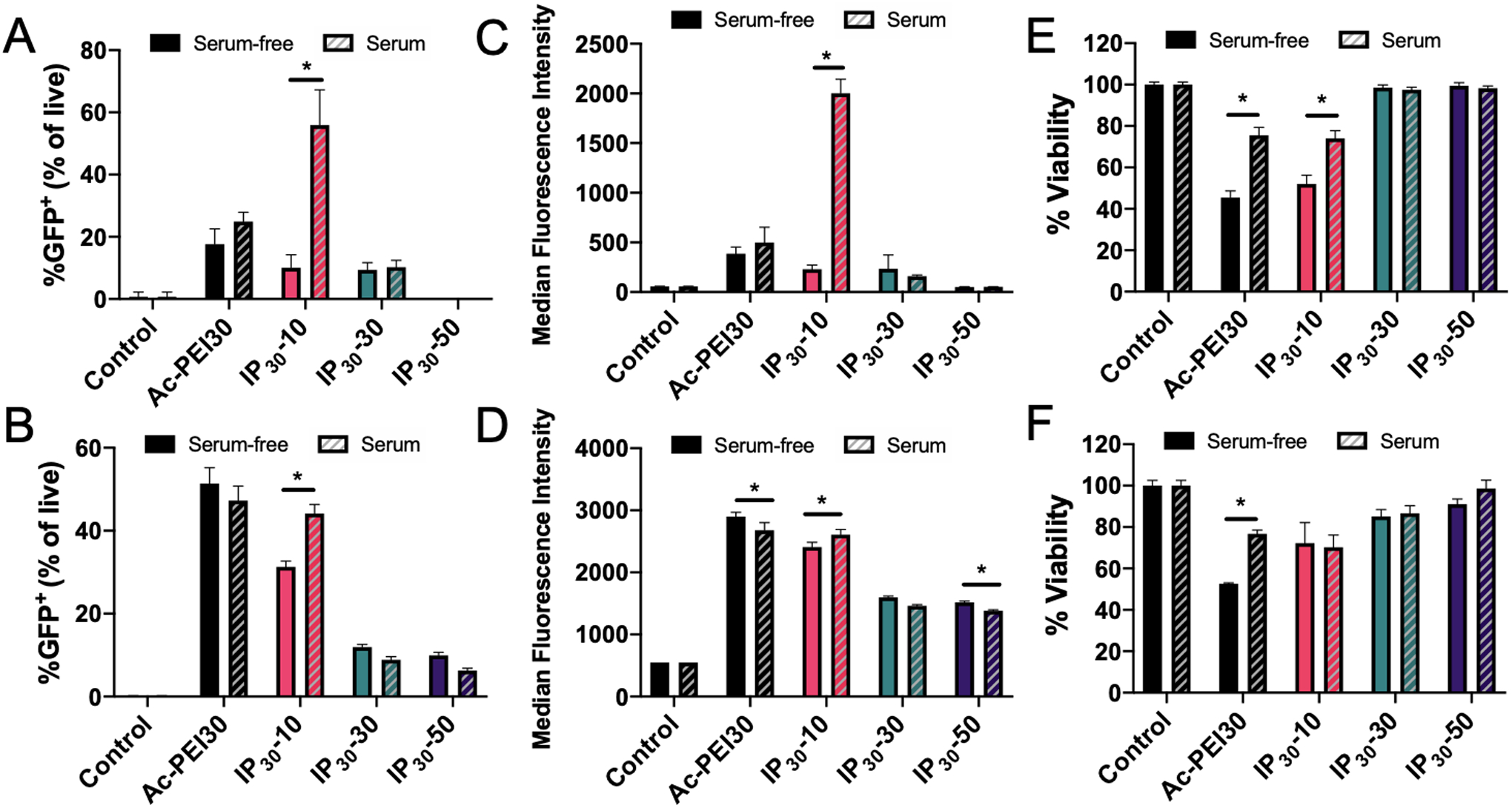
Transfection efficiency of IPs prepared with various wt.% of PEMA (10% to 50%) determined by flow cytometry in RAW 264.7 (A) and Jurkat cells (B). Median Fluorescence Intensity of GFP expression for RAW 264.7 (C) and Jurkat cells (D). Cell viability determined by MTS assay for RAW 264.7 (E) and Jurkat cells (F). Cells were incubated in serum-free or serum-containing medium for 4 hrs with polyplexes, washed, and incubated for 24 hrs prior to analysis. Data are representative of n=3 experiments. Statistical differences between groups were determined by performing a one-way ANOVA and Tukey’s post hoc test (p < 0.05). Error bars represent SD.