
Figure 3. SFL Interacts with Ribosomes.
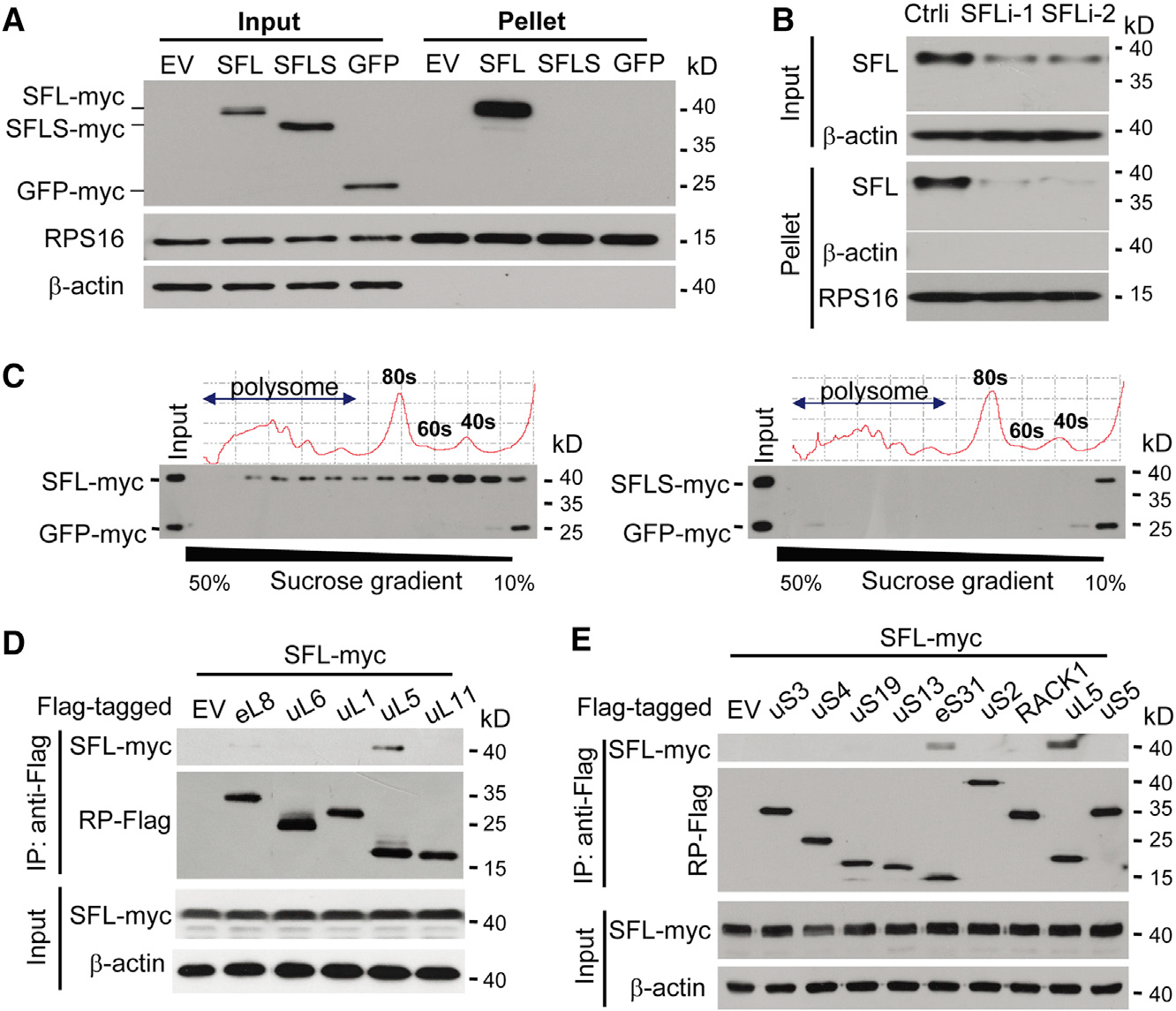
(A) Ribosomes in the lysates of 293T cells transiently expressing SFL-Myc, SFLS-Myc, or GFP-Myc were pelleted and analyzed by western blotting. RPS16 is an endogenous ribosomal protein.
(B) HeLa cells were transfected with a control siRNA or siRNAs targeting SFL. Ribosomes were pelleted and analyzed by western blotting.
(C) SFL-Myc (left) or SFLS-Myc (right) was transiently expressed in 293T cells with GFP-Myc. The cell lysates were subjected to sucrose gradient centrifugation, followed by polysome profiling analysis. Protein levels in each fraction were analyzed by western blotting.
(D and E) Interactions of SFL-Myc with Flag-tagged ribosome large-subunit (D) and small-subunit (E) proteins in 293T cells.
See also Figure S3.