
Figure 7. Working Model of SFL.
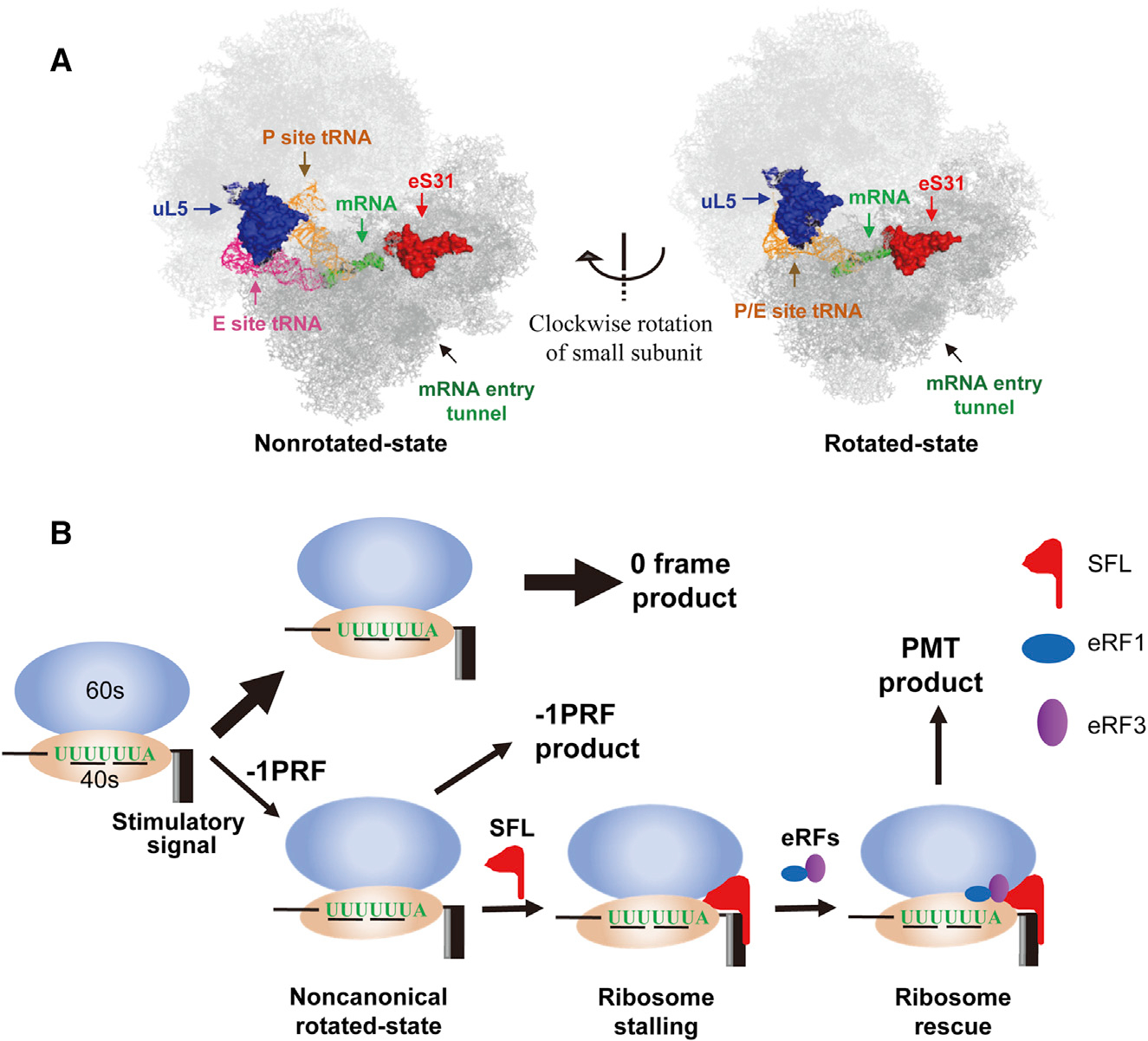
(A) Positions of uL5, eS31, and mRNA entry tunnel in the nonrotated state (left) and rotated state (right) of the ribosome. The structures are adapted from a previously published paper (Svidritskiy et al., 2014).
(B) A working model for SFL inhibition of −1PRF. When the translating ribosome is in a noncanonical rotated state during the process of −1PRF, SFL stably binds to the ribosome, causing ribosome stalling. The eRF1-eRF3 complex is recruited to rescue the stalled ribosome, leading to the production of PMT product.
See also Figure S7.