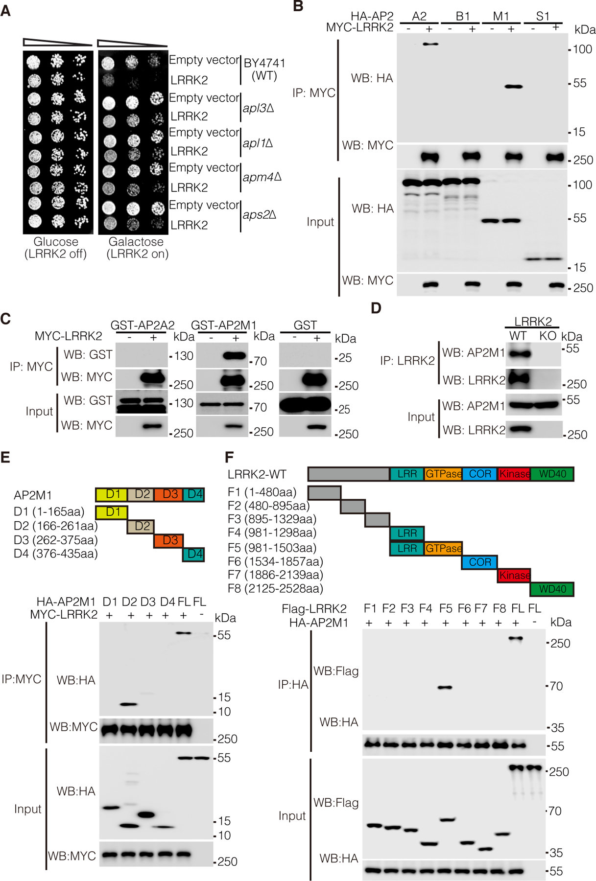
Figure 1. AP2M1 interacts with LRRK2.

(A) Cell viability assay by cell number quantitation in response to expression of GTP-COR-Kin fragment of WT LRRK2 (pYES2-LRRK2-GCK) or empty vector in WT and AP2 subunit-deleted (apl3∆, apl1∆, apm4∆, aps2∆) BY4741 yeast strains. Cells were spotted onto media containing glucose (LRRK2 Off, repressed, left panel) or galactose (LRRK2 On, induced, right panel) and incubated at 30°C for 2 to 3 days. Shown are five-fold serial dilutions of yeast cells from left to right as indicated by graded open box. (B) Coimmunoprecipitation (co-IP) analysis for interaction between MYC-tagged LRRK2 and HA-tagged AP2 subunits (A2, B1, M1, or S1) in co-transfected HEK 293T cells. Co-IP with antibody to MYC was followed by immunoblotting for HA or MYC. (C) Co-IP analysis of the interactions between AP2 subunits (A2, M1) and LRRK2 using recombinant GST-tagged AP2M1 and A2 subunits and purified MYC-LRRK2. Co-IP with antibody to MYC was followed by immunoblotting for GST. GST protein was as a negative control. (D) Co-IP analysis of the interactions between AP2M1 and LRRK2 in mouse brain lysates. Whole brain lysates prepared form wild-type (WT) and LRRK2 knockout (KO) mice were subjected to co-IP with antibody to LRRK2 (Neuromab) followed by immunoblotting for LRRK2 and AP2M1. (E) Co-IP analysis of the interactions between MYC-LRRK2 and HA-tagged D1, D2, D3, D4 or full-length (FL) AP2M1 in co-transfected HEK 293T cells. Co-IP with antibody to MYC was followed by immunoblotting for HA or MYC. A schematic representation of AP2M1 D1, D2, D3, D4 domains is shown. (F) Co-IP analysis of the interactions between HA-tagged AP2M1 and Flag-tagged LRRK2 fragments in co-transfected HEK 293T cells. Co-IP with antibody to HA was followed by immunoblotting for Flag or HA. A schematic representation of the LRRK2 fragments is shown. Blots are representative of 3 experiments.
