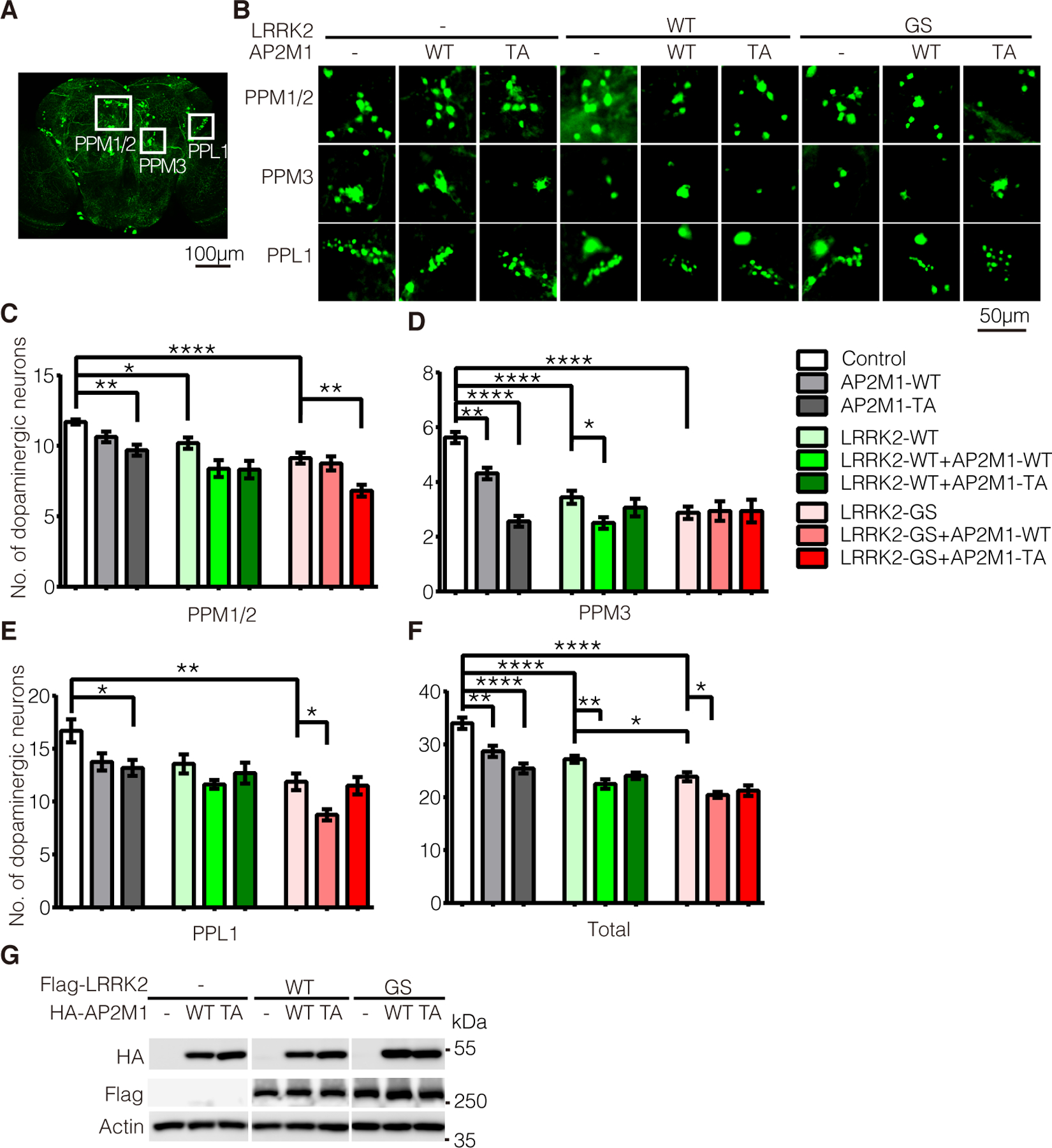
Figure 7. LRRK2 phosphorylation of AP2M1 modulates LRRK2-induced dopaminergic neurodegeneration in vivo.

(A) Diagram of dopaminergic neuronal clusters (PPM1/2, PPM3, PPL1) in the posterior areas of the adult fly brain. Scale bars, 100 μm. (B) Representative confocal images (GFP) of dopamine neurons in each dopaminergic cluster from 7-week-old flies of the indicated genotypes. Scale bars, 50 μm. (C to E) Quantification of dopamine neurons per dopaminergic cluster in 7-weeks old flies of the indicated genotypes. (F) Total numbers of dopamine neurons in four major dopaminergic clusters of the flies at 7-weeks old. Data are the means ± SEM, n=8 flies per genotype, *P < 0.05, **P < 0.01, ***P < 0.001, and ****P < 0.0001 by one-way ANOVA followed by a Tukey’s post hoc test. (G) Levels of overexpressed LRRK2 and AP2M1 in flies driven by GMR-Gal4. Lysates prepared form whole heads of one-week old flies of the indicated genotypes were subjected to immunoblotting with antibodies to HA-HRP, Flag-HRP and fly actin. Blots are representative of n=3 independent experiments.
